You have no items in your shopping cart.
Featured
Description
Research Area
Cancer Biology
Images & Validation
−Item 1 of 5
| Tested Applications | FC, IF, IHC-P, WB |
|---|---|
| Reactivity | Human |
| Application Notes |
Key Properties
−| Antibody Type | Primary Antibody |
|---|---|
| Host | Rabbit |
| Clonality | Polyclonal |
| Isotype | Rabbit Ig |
| Immunogen | This TIMP1 antibody is generated from rabbits immunized with a KLH conjugated synthetic peptide between 157-188 amino acids from the C-terminal region of human TIMP1. |
| Target | TIMP1 |
| Molecular Weight | 23 kDa |
| Purification | This antibody is prepared by Saturated Ammonium Sulfate (SAS) precipitation followed by dialysis |
| Conjugation | Unconjugated |
Storage & Handling
−| Storage | Maintain refrigerated at 2-8°C for up to 2 weeks. For long term storage store at -20°C in small aliquots to prevent freeze-thaw cycles. |
|---|---|
| Form/Appearance | Liquid |
| Buffer/Preservatives | Supplied in PBS with 0.09% (W/V) sodium azide. |
| Concentration | batch dependent |
| Expiration Date | 12 months from date of receipt. |
| Disclaimer | For research use only |
Alternative Names
−Metalloproteinase inhibitor 1, Erythroid-potentiating activity, EPA, Fibroblast collagenase inhibitor, Collagenase inhibitor, Tissue inhibitor of metalloproteinases 1, TIMP-1, TIMP1, CLGI, TIMP
Similar Products
−- Item 1 of 18
TIMP1 Rabbit Polyclonal Antibody [orb195994]
ELISA, ICC, IF, IHC-P, WB
Bovine, Canine, Guinea pig, Human, Mouse, Porcine, Rat, Sheep
Rabbit
Polyclonal
Unconjugated
100 μg - Item 1 of 10
TIMP-1 Mouse Monoclonal Antibody [orb500828]
FC, ICC
Mouse, Rat
Human
Mouse
Monoclonal
Unconjugated
50 μl, 100 μl, 200 μl, 200 μg - Item 1 of 7
TIMP1 Rabbit Polyclonal Antibody [orb11483]
ELISA, ICC, IF, IHC-P, WB
Rabbit
Polyclonal
Unconjugated
100 μg - Item 1 of 5
TIMP-1 Rabbit Polyclonal Antibody [orb100174]
WB
Bovine, Canine, Mouse, Porcine, Rabbit, Sheep
Human
Rabbit
Polyclonal
Unconjugated
100 μl, 200 μl, 50 μl - Item 1 of 5
TIMP-1 Rabbit Polyclonal Antibody [orb313247]
WB
Bovine, Canine, Porcine, Rat, Sheep
Human
Rabbit
Polyclonal
Unconjugated
50 μl, 100 μl, 200 μl
Quality Guarantee
Explore bioreagents carefree to elevate your research. All our products are rigorously tested for performance. If a product does not perform as described on its datasheet, our scientific support team will provide expert troubleshooting, a prompt replacement, or a refund. For full details, please see our Terms & Conditions and Buying Guide. Contact us at [email protected].
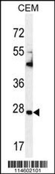
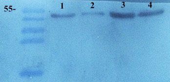
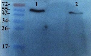
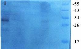
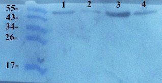
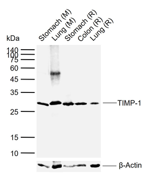
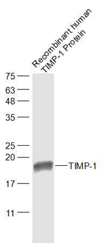
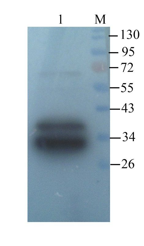
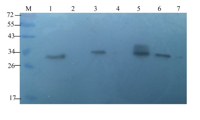
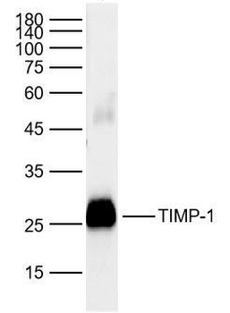
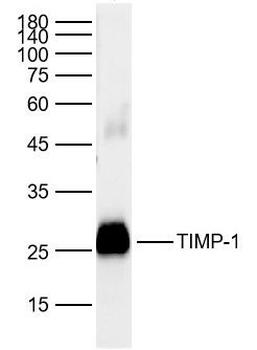
Western blot analysis in CEM cell line lysates (35 ug/lane).
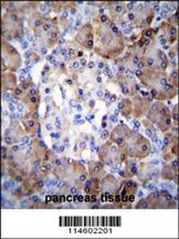
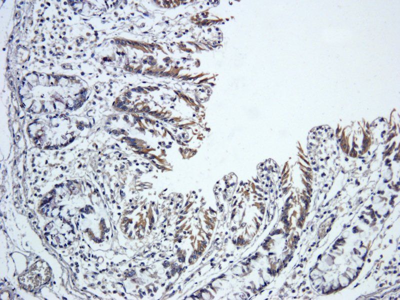
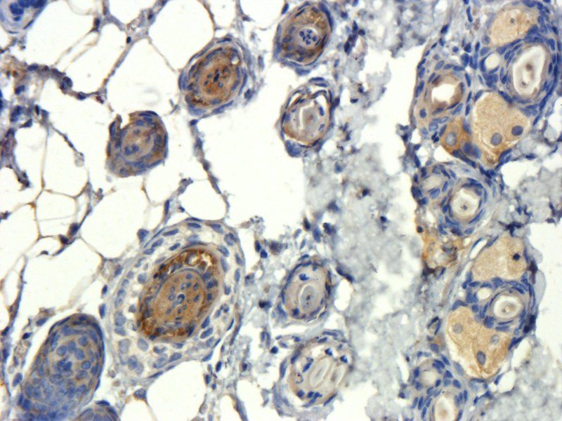
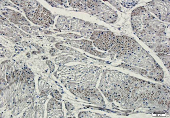
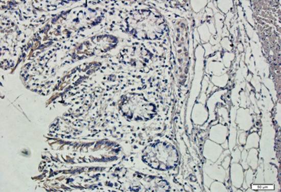
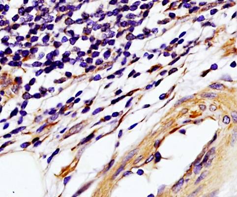
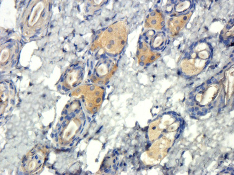
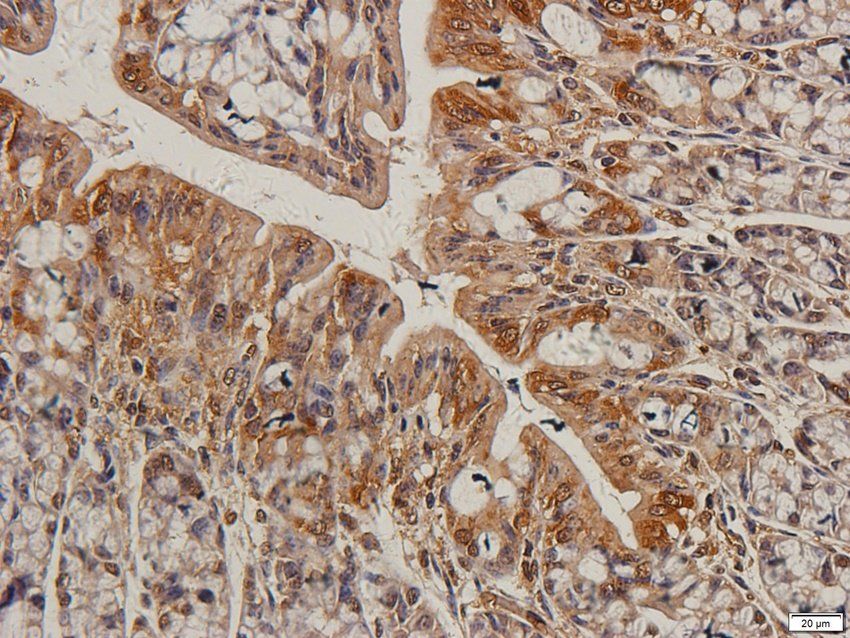
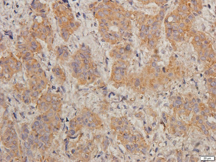
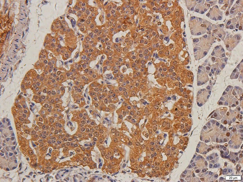
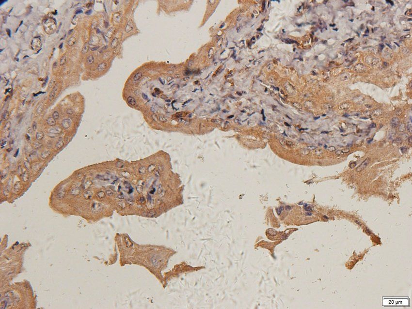
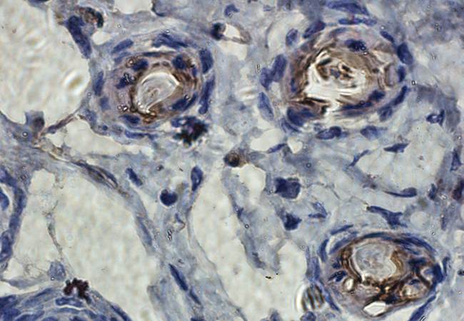
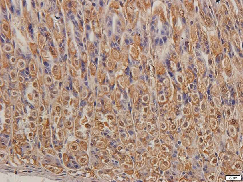
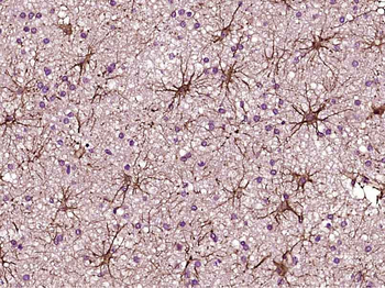
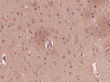
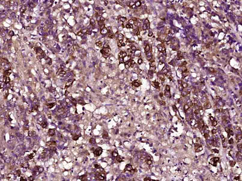
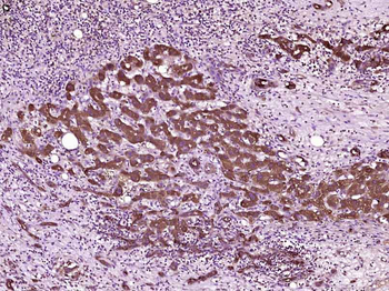
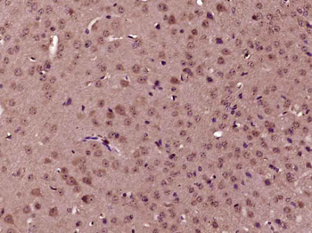
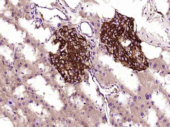
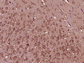
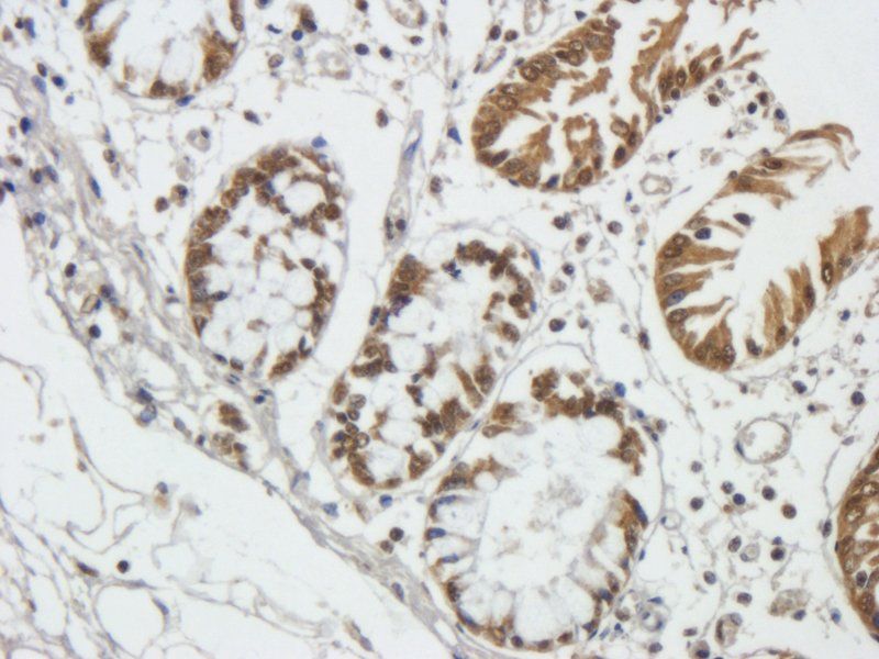
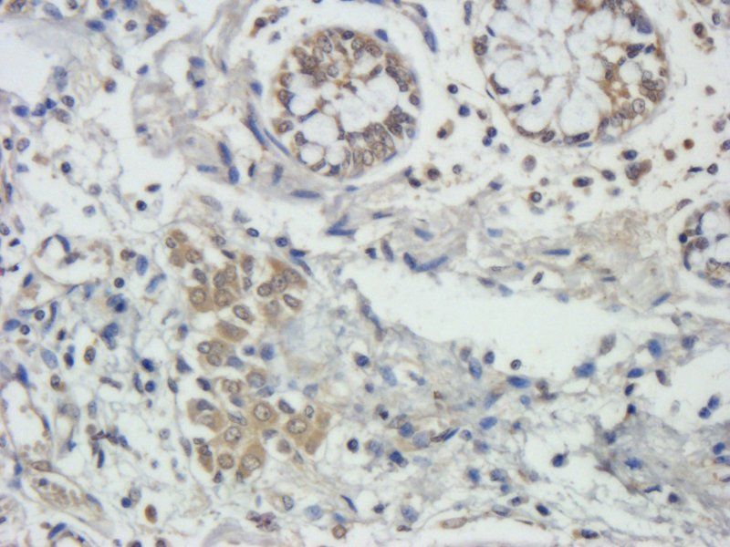
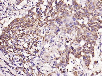
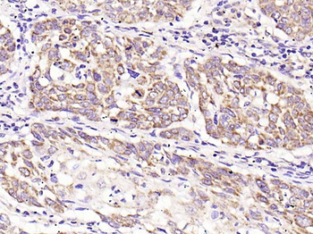
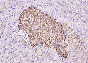
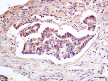
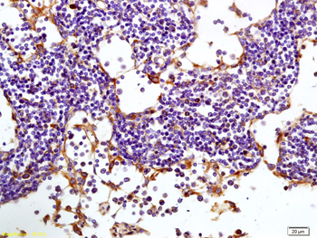
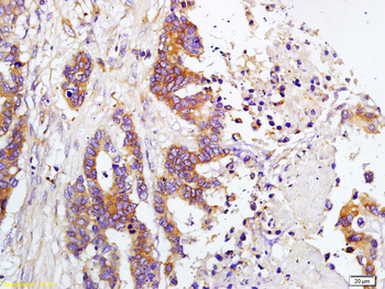
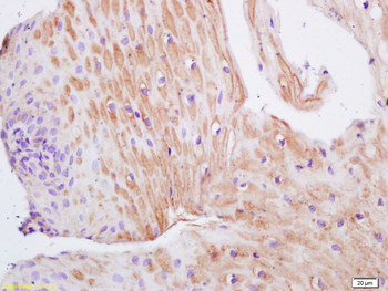
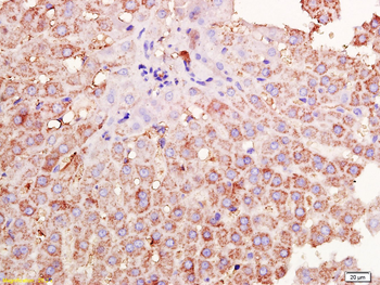
TIMP1 Antibody immunohistochemistry analysis in formalin fixed and paraffin embedded human pancreas tissue followed by peroxidase conjugation of the secondary antibody and DAB staining.
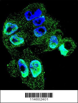
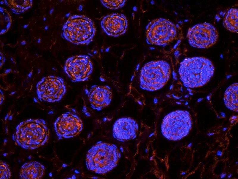
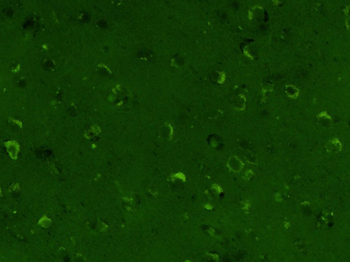
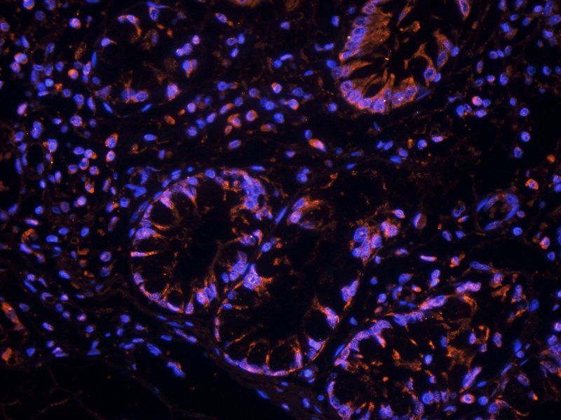
Confocal immunofluorescent analysis of TIMP1 Antibody with A2058 cell followed by Alexa Fluor 488-conjugated goat anti-rabbit lgG (green). DAPI was used to stain the cell nuclear (blue).
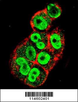
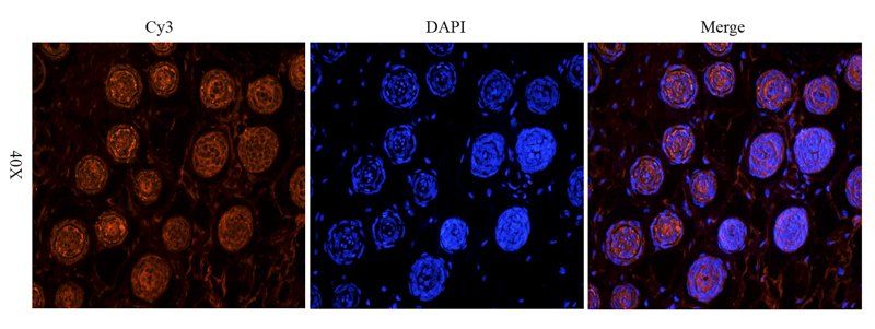
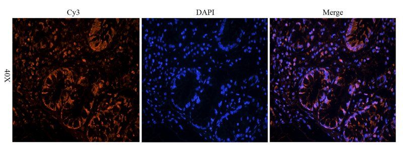
Confocal immunofluorescent analysis of TIMP1 Antibody with A2058 cell followed by Alexa Fluor 488-conjugated goat anti-rabbit lgG (green). Actin filaments have been labeled with Alexa Fluor 555 phalloidin (red).
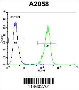
Flow cytometric analysis of A2058 cells (right histogram) compared to a negative control cell (left histogram). FITC-conjugated goat-anti-rabbit secondary antibodies were used for the analysis.
Documents Download
Datasheet
Product Information
Request a Document
Protocol Information
WB
Western Blot (IB, immunoblot)
IHC-P
Immunohistochemistry Paraffin
FC
Flow Cytometry
IF
Immunofluorescence
TIMP1 Antibody (orb1270310)

- 0.0
Based on 0 reviews
Participating in our Biorbyt product reviews program enables you to support fellow scientists by sharing your firsthand experience with our products.
Login to Submit a ReviewAvailable Sizes
Select a size below
Free Secondary Antibody (20 ul)0/0
Please add an antibody product to your cart first.