You have no items in your shopping cart.
Description
Research Area
Infectious Disease & Virology, Neuroscience
Images & Validation
−Item 1 of 6
| Tested Applications | IHC, WB |
|---|---|
| Dilution Range | WB: 1:2000, WB: 1:3000, IHC: 1:25, IHC: 1:25, IHC: 1:25, IHC: 1:25 |
| Reactivity | Human, Mouse |
Key Properties
−| Antibody Type | Primary Antibody |
|---|---|
| Host | Mouse |
| Clonality | Monoclonal |
| Isotype | IgG1 |
| Immunogen | Purified His-tagged TBP protein was used to produced this monoclonal antibody. Antigen Region: 1-288 aa. |
| Target | TBP |
| Molecular Weight | 37698 Da |
| Conjugation | Unconjugated |
Storage & Handling
−| Storage | Maintain refrigerated at 2-8°C for up to 2 weeks. For long term storage store at -20°C in small aliquots to prevent freeze-thaw cycles |
|---|---|
| Expiration Date | 12 months from date of receipt. |
| Disclaimer | For research use only |
Alternative Names
−TATA-box-binding protein, TATA sequence-binding protein, TATA-binding factor, TATA-box factor, Transcription initiation factor TFIID TBP subunit, TBP, GTF2D1, TF2D, TFIID
Similar Products
−- Item 1 of 13
TAF4 Rabbit Polyclonal Antibody [orb745927]
ELISA, ICC, IF, IHC, WB
Human, Mouse, Rat
Rabbit
Polyclonal
Unconjugated
100 μg - Item 1 of 10
TIP49A/RUVBL1 Rabbit Polyclonal Antibody [orb1173474]
ELISA, FC, ICC, IF, IHC, IP, WB
Human, Monkey, Mouse, Rat
Rabbit
Polyclonal
Unconjugated
100 μg - Item 1 of 7
TBP-1/PSMC3/TBP Rabbit Polyclonal Antibody [orb670321]
ELISA, FC, IHC, WB
Human, Mouse, Rat
Rabbit
Polyclonal
Unconjugated
100 μg - Item 1 of 6
- Item 1 of 4
TBP Antibody (Center) [orb1929890]
FC, IF, IHC-P, WB
Mouse, Other, Zebrafish
Human
Rabbit
Polyclonal
Unconjugated
100 μl, 50 μl


Quality Guarantee
Explore bioreagents carefree to elevate your research. All our products are rigorously tested for performance. If a product does not perform as described on its datasheet, our scientific support team will provide expert troubleshooting, a prompt replacement, or a refund. For full details, please see our Terms & Conditions and Buying Guide. Contact us at [email protected].
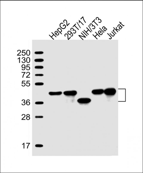
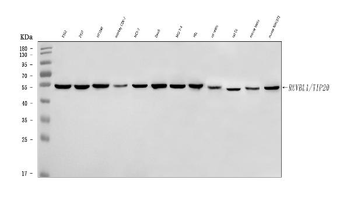
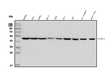
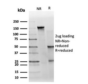
All lanes: Anti-TBP Antibody at 1:2000 dilution. Lane 1: HepG2 whole cell lysate. Lane 2: 293T/17 whole cell lysate. Lane 3: NIH/3T3 whole cell lysate. Lane 4: Hela whole cell lysate. Lane 5: Jurkat whole cell lysate. Lysates/proteins at 20 µg per lane. Secondary Goat Anti-mouse IgG, (H+L), Peroxidase conjugated at 1/10000 dilution. Predicted band size: 38 kDa. Blocking/Dilution buffer: 5% NFDM/TBST.
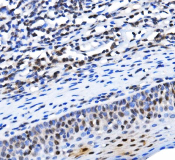
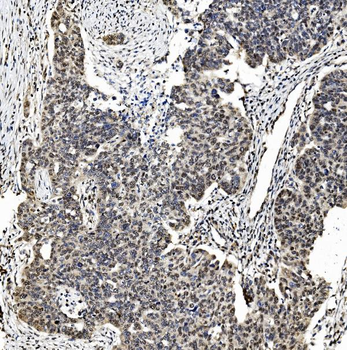
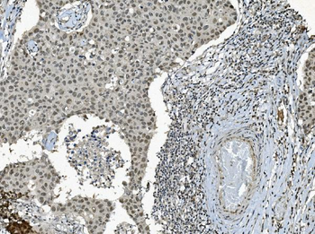
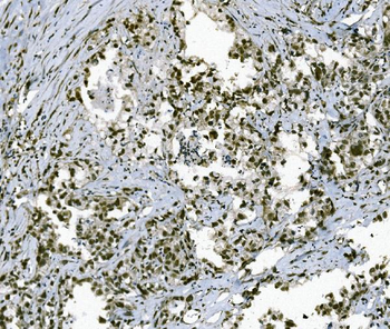
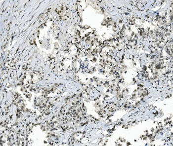
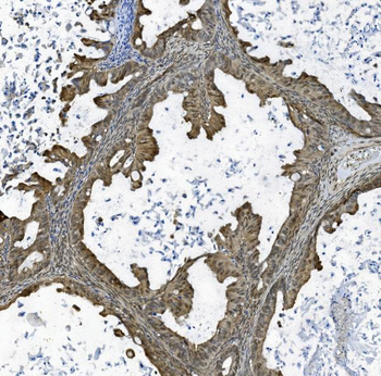
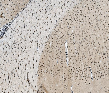
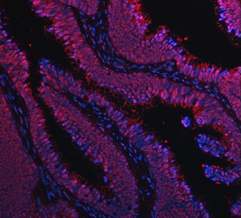
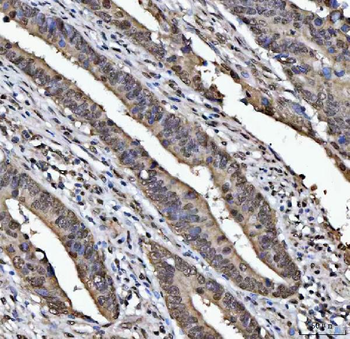
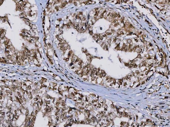
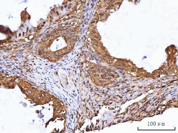
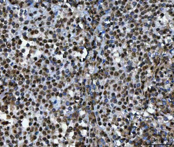
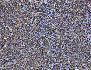
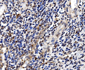
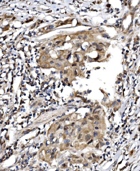
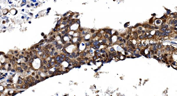
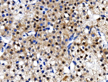
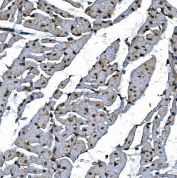
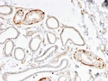
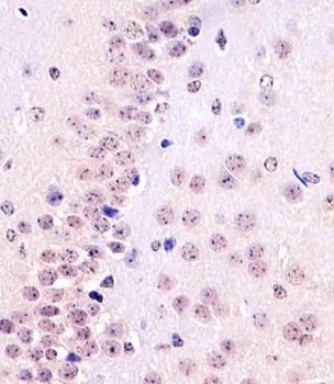
Staining TBP in Rat brain tissue sections by Immunohistochemistry (IHC-P - paraformaldehyde-fixed, paraffin-embedded sections). Tissue was fixed with formaldehyde and blocked with 3% BSA for 0.5 hour at room temperature; antigen retrieval was by heat mediation with a citrate buffer (pH6). Samples were incubated with primary antibody (1/25) for 1 hours at 37°C. A undiluted biotinylated goat polyvalent antibody was used as the secondary antibody.
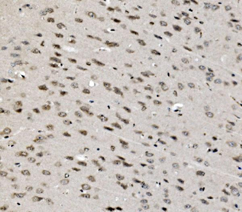
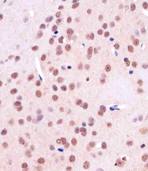
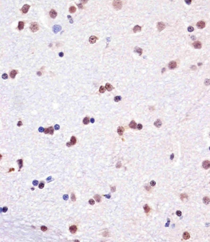
Staining TBP in mouse brain tissue sections by Immunohistochemistry (IHC-P - paraformaldehyde-fixed, paraffin-embedded sections). Tissue was fixed with formaldehyde and blocked with 3% BSA for 0.5 hour at room temperature; antigen retrieval was by heat mediation with a citrate buffer (pH6). Samples were incubated with primary antibody (1/25) for 1 hours at 37°C. A undiluted biotinylated goat polyvalent antibody was used as the secondary antibody.
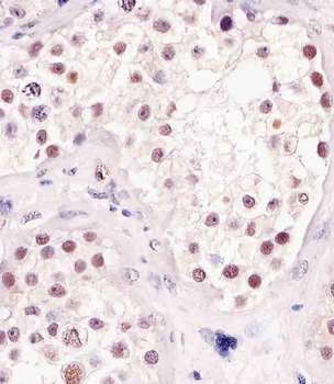
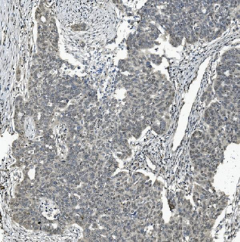
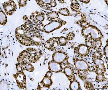
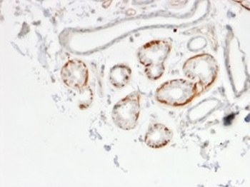
Staining TBP in human testis tissue sections by Immunohistochemistry (IHC-P - paraformaldehyde-fixed, paraffin-embedded sections). Tissue was fixed with formaldehyde and blocked with 3% BSA for 0.5 hour at room temperature; antigen retrieval was by heat mediation with a citrate buffer (pH6). Samples were incubated with primary antibody (1/25) for 1 hours at 37°C. A undiluted biotinylated goat polyvalent antibody was used as the secondary antibody.
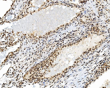
Staining TBP in Monkey. brain tissue sections by Immunohistochemistry (IHC-P - paraformaldehyde-fixed, paraffin-embedded sections). Tissue was fixed with formaldehyde and blocked with 3% BSA for 0.5 hour at room temperature; antigen retrieval was by heat mediation with a citrate buffer (pH6). Samples were incubated with primary antibody (1/25) for 1 hours at 37°C. A undiluted biotinylated goat polyvalent antibody was used as the secondary antibody.
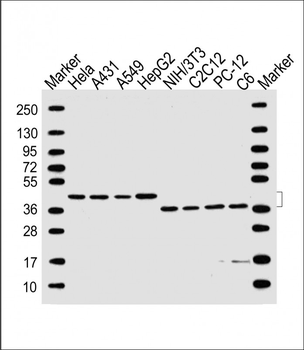
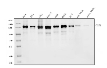
All lanes: Anti-TBP Antibody at 1: 3000 dilution. Lane 1: Hela whole cell lysate. Lane 2: A431 whole cell lysate. Lane 3: A549 whole cell lysate. Lane 4: HepG2 whole cell lysate. Lane 5: NIH/3T3 whole cell lysate. Lane 6: C2C12 whole cell lysate. Lane 7: PC-12 whole cell lysate. Lane 8: C6 whole cell lysate. Lysates/proteins at 20 µg per lane. Secondary Goat Anti-mouse IgG, (H+L), Peroxidase conjugated at 1/10000 dilution. Predicted band size: 38 kDa. Blocking/Dilution buffer: 5% NFDM/TBST.
Quick Database Links
UniProt Details
− No UniProt data available
NCBI Reference Sequences
−Associated Accession Numbers
Curated reference sequences for the gene transcript and protein product| Protein | NP_001165556.1, NP_003185.1 |
|---|
Documents Download
Datasheet
Product Information
Request a Document
Protocol Information
WB
Western Blot (IB, immunoblot)
IHC
Immunohistochemistry
TBP Antibody (orb1787877)

- 0.0
Based on 0 reviews
Participating in our Biorbyt product reviews program enables you to support fellow scientists by sharing your firsthand experience with our products.
Login to Submit a Review