You have no items in your shopping cart.
Featured
Description
Research Area
Neuroscience
Images & Validation
−Item 1 of 5
| Tested Applications | DOT, ELISA, ICC, IF, IHC, WB |
|---|---|
| Dilution Range | WB (1:500), ICC/IF (1:500); IHC (1:500) |
| Reactivity | Human, Mouse |
| Application Notes |
Key Properties
−| Host | Rabbit |
|---|---|
| Clonality | Monoclonal |
| Isotype | IgG |
| Clone No. | AH36 |
| Immunogen | Synthetic peptide of Human Phospho Tau (Ser202/Thr205) |
| Target | Tau (pSer202/ pThr205) |
| Molecular Weight | 79 kDa |
| Purification | Affinity Purified |
| Conjugation | Unconjugated |
Storage & Handling
−| Storage | Maintain refrigerated at 2-8°C for up to 2 weeks. For long term storage store at -20°C in small aliquots to prevent freeze-thaw cycles. |
|---|---|
| Buffer/Preservatives | PBS pH 7.4, 50% glycerol, 0.09% sodium azide. Storage buffer changes when conjugated. |
| Concentration | 1 mg/ml |
| Expiration Date | 12 months from date of receipt. |
| Disclaimer | For research use only |
Alternative Names
−Tau, TAU, TAU_HUMAN, MAPT, MAPTL, Microtubule-associated protein tau, Microtubule associated protein tau, Microtubule associated protein tau isoform 4, Neurofibrillary tangle protein, Paired helical filament tau, Paired helical filament-tau, PHF tau, PHF-tau, DDPAC, FTDP 17, G protein beta1/gamma2 subunit interacting factor 1, MSTD, Mtapt, MTBT1, MTBT2, PPND, PPP1R103, Protein phosphatase 1 regulatory subunit 103, RNPTAU, AI413597, AW045860, FLJ31424, MGC134287, MGC138549, MGC156665
Similar Products
−- Item 1 of 5
Tau (pSer202/ pThr205) Antibody (APC) [orb612742]
DOT, ELISA, ICC, IF, IHC, WB
Human, Mouse
Rabbit
Monoclonal
APC
100 μg - Item 1 of 5
Tau (pSer202/ pThr205) Antibody (Biotin) [orb612744]
DOT, ELISA, ICC, IF, IHC, WB
Human, Mouse
Rabbit
Monoclonal
Biotin
100 μg - Item 1 of 5
Tau (pSer202/ pThr205) Antibody (FITC) [orb612745]
DOT, ELISA, ICC, IF, IHC, WB
Human, Mouse
Rabbit
Monoclonal
FITC
100 μg - Item 1 of 5
Tau (pSer202/ pThr205) Antibody (HRP) [orb612746]
DOT, ELISA, ICC, IF, IHC, WB
Human, Mouse
Rabbit
Monoclonal
HRP
100 μg - Item 1 of 5
Tau (pSer202/ pThr205) Antibody (PerCP) [orb612748]
DOT, ELISA, ICC, IF, IHC, WB
Human, Mouse
Rabbit
Monoclonal
PerCP
100 μg


Quality Guarantee
Explore bioreagents carefree to elevate your research. All our products are rigorously tested for performance. If a product does not perform as described on its datasheet, our scientific support team will provide expert troubleshooting, a prompt replacement, or a refund. For full details, please see our Terms & Conditions and Buying Guide. Contact us at [email protected].

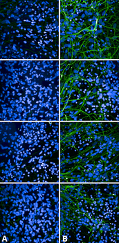
Immunocytochemistry/Immunofluorescence analysis using Rabbit Anti-Tau Monoclonal Antibody, Clone AH36. Tissue: iPSC-derived cortical excitatory neurons. Species: Human. Primary Antibody: Rabbit Anti-Tau Monoclonal Antibody at 1:500 for Overnight. Secondary Antibody: Donkey anti-rabbit: Alexa Fluor 488 at 1:1000. Counterstain: DAPI. A) iPSC-derived neurons from non-demented control (NDC). B) iPSC-derived neurons from subject with P301L MAPT mutation. Images acquired using an automated Opera Phoenix system. Each field of view is a max projection from 10 planes of 1 μm stacks.

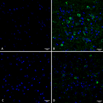
Immunohistochemistry analysis using Rabbit Anti-Tau Monoclonal Antibody, Clone AH36. Tissue: Brain slice. Species: Mouse. Primary Antibody: Rabbit Anti-Tau Monoclonal Antibody at 1:500 for Overnight at 4°C. Secondary Antibody: Anti-Rabbit IgG: AlexaFluor 488. Counterstain: DAPI at 1:1000 for 5 min. (A) Pons of Non-Tg mouse. (B) Pons of P301SxUBQLN2 Tg mouse. (C) Prefrontal cortex of Non-Tg mouse. (D) Prefrontal cortex of P301SxUBQLN2 Tg mouse. IHC Protocol: 1. Post-fix brains in 4% PFA for 24 hours and put through a 10-30% sucrose gradient. 2. Section by cryostat at 10 uM thickness. 3. Fix in MeOH 15 min. 4. 3x10 min wash in PBS 1X. 5. Heat via microwave in 10mM Citrate Buffer, pH 6 for 4 min at power level 20. 6. Cool in solution for 20 min. 7. Wash 2x5 min in PBS. 8. Permeabilize in 0.5% Triton-X 100 in PBS 10 min. 9. Wash in PBS 10 min. 10. Block for 1 hour in 5% goat serum. 11. Incubate primary Ab (at 1:500) in blocking solution overnight at 4°C. 12. Wash 3x10 min in PBS. 13. Incubate in secondary Ab Rb IgG Alexa-fluor 488. 14. Wash 3x10 min in PBS. 15. Incubate in DAPI 1:1000 for 5 min. 16. Wash 3x5 min. 17. Coverslip with Prolong-Gold.
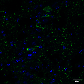
Immunohistochemistry analysis using Rabbit Anti-Tau Monoclonal Antibody, Clone AH36. Tissue: Brain slice. Species: Mouse. Primary Antibody: Rabbit Anti-Tau Monoclonal Antibody at 1:500 for Overnight at 4°C. Secondary Antibody: Anti-Rabbit IgG: AlexaFluor 488. Counterstain: DAPI at 1:1000 for 5 min. CA3 Region of P301SxUBQLN2 Tg mouse. IHC Protocol: 1. Post-fix brains in 4% PFA for 24 hours and put through a 10-30% sucrose gradient. 2. Section by cryostat at 10 uM thickness. 3. Fix in MeOH 15 min. 4. 3x10 min wash in PBS 1X. 5. Heat via microwave in 10mM Citrate Buffer, pH 6 for 4 min at power level 20. 6. Cool in solution for 20 min. 7. Wash 2x5 min in PBS. 8. Permeabilize in 0.5% Triton-X 100 in PBS 10 min. 9. Wash in PBS 10 min. 10. Block for 1 hour in 5% goat serum. 11. Incubate primary Ab (at 1:500) in blocking solution overnight at 4°C. 12. Wash 3x10 min in PBS. 13. Incubate in secondary Ab Rb IgG Alexa-fluor 488. 14. Wash 3x10 min in PBS. 15. Incubate in DAPI 1:1000 for 5 min. 16. Wash 3x5 min. 17. Coverslip with Prolong-Gold.

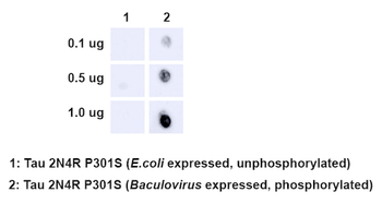
Dot Blot analysis using Rabbit Anti-Tau Monoclonal Antibody, Clone AH36. Species: E. Coli, Baculovirus. Primary Antibody: Rabbit Anti-Tau Monoclonal Antibody at 1:500. Secondary Antibody: Goat anti-rabbit IgG:HRP.

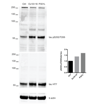
Western Blot analysis of Human iPSC-derived cortical neurons showing detection of Tau protein using Rabbit Anti-Tau Monoclonal Antibody, Clone AH36. Lane 1: MW ladder. Lane 2: Control (non-disease) line. Lane 2: Ex10+16 tau mutant sample. Lane 3: P301L tau mutant sample. Load: 50ug. Primary Antibody: Rabbit Anti-Tau Monoclonal Antibody at 1:500 for Overnight. Total tau was detected using mouse anti-tau antibody (clone HT7). The bar graph on the right shows quantification of pSer202/pThr205 compared to total tau in each sample.
Documents Download
Datasheet
Product Information
Request a Document
Protocol Information
WB
Western Blot (IB, immunoblot)
IHC
Immunohistochemistry
IF
Immunofluorescence
ICC
Immunocytochemistry
ELISA
Enzyme-linked Immunosorbent Assay (EIA)
DOT
Dot Blot
Tau (pSer202/ pThr205) Antibody (orb612732)

- 0.0
Based on 0 reviews
Participating in our Biorbyt product reviews program enables you to support fellow scientists by sharing your firsthand experience with our products.
Login to Submit a ReviewAvailable Sizes
Select a size below
Choose Conjugation or Carrier Free Version
Free Secondary Antibody (20 ul)0/0
Please add an antibody product to your cart first.