You have no items in your shopping cart.
Streptavidin
SKU: orb348765
Description
Research Area
Non-Animal
Images & Validation
−Item 1 of 7
| Application Notes |
|---|
Key Properties
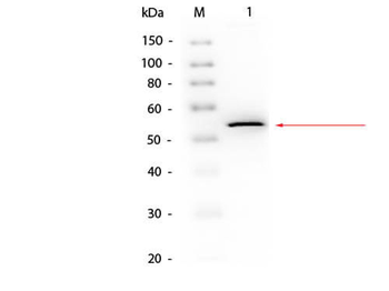
−| Purity | Streptavidin is chromatographically pure and shows predominantly a single 53,600 dalton band by SDS-PAGE. |
|---|---|
| Biological Activity | 15.5 U/mg by biotin titration method |
| Conjugation | Unconjugated |
Storage & Handling
−| Storage | Store vial at 4° C prior to restoration. For extended storage aliquot contents and freeze at -20° C or below. Avoid cycles of freezing and thawing. Centrifuge product if not completely clear after standing at room temperature. Streptavidin is stable for several weeks at 4° C as an undiluted liquid. Dilute only prior to immediate use. |
|---|---|
| Form/Appearance | Lyophilized |
| Buffer/Preservatives | Preservative: None. Stabilizer: None; Buffer: 0.15 M Sodium Chloride |
| Concentration | 0.9 mg/mg |
| Expiration Date | 12 months from date of receipt. |
| Hazard Information | Non-Toxic |
| Disclaimer | For research use only |
Alternative Names
−SA, S avidin, streptococcus avidin
Similar Products
−- Item 1 of 9
- Item 1 of 10
- Item 1 of 2

ELISA Pro: Human IL-6 [orb1532209]
ELISA

Rabbit Mouse IgE (Fc specific), conjugated with Biotin Antibody [orb21623]
Mouse
Rabbit
Polyclonal
Biotin
1 ml

Quality Guarantee
Explore bioreagents carefree to elevate your research. All our products are rigorously tested for performance. If a product does not perform as described on its datasheet, our scientific support team will provide expert troubleshooting, a prompt replacement, or a refund. For full details, please see our Terms & Conditions and Buying Guide. Contact us at [email protected].
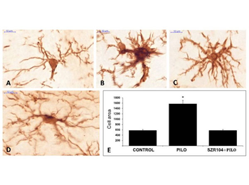
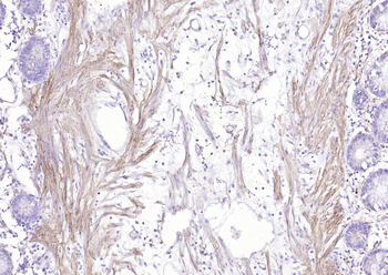
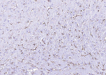
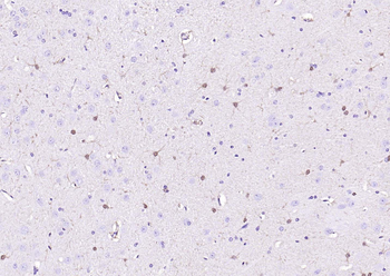
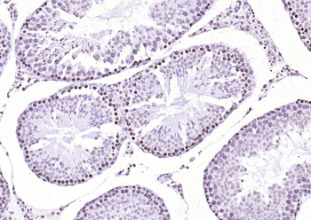
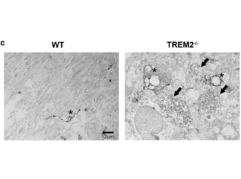
(A–D): ionized calcium-binding adaptor molecule 1 (Iba1)-stained microglia cells from control (A), pilocarpine(PILO)-treated (B, C) and SZR104 + PILO-treated (D) animals. Scale bars: 10 µm. (E): The average microglia cell areas (cell body and processes) in µm2 values on the y-axis. Biotinylated secondary antibodies (1:400) and the signal was detected with peroxidase-labeled streptavidin (1:6000) (p/n orb348765). (n = 10; mean ± SEM) in control, PILO-treated and SZR104 + PILO-treated animals (* p ≤ 0.05).
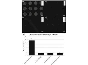
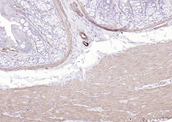
Development of PDMS MB well surface coating to enhance capture cells with affinity for α-IgG antibody. FITC-conjugated IgG was added to MB wells coated with a streptavidin + biotinylated α-IgG, b biotinylated α-IgG only, c uncoated MBs and d streptavidin + biotinylated α-Transferrin. Quantification of the fluorescent intensity averaged over 12 wells is illustrated in (e). Results indicate that FITC-IgG predominantly binds to the (SA) + b-α(IgG) coating. Bars indicate standard error, n = 12, *P < 0.0001. Streptavidin (p/n orb348765).
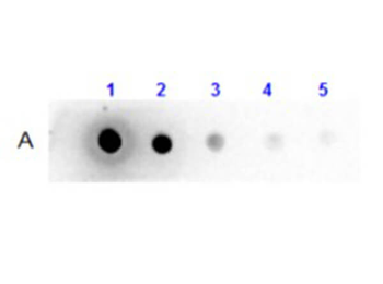
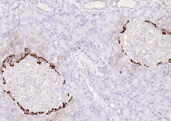
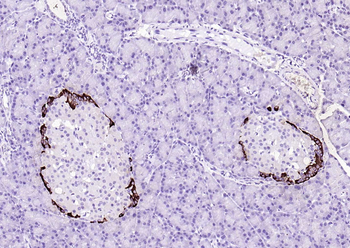
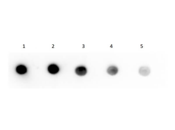
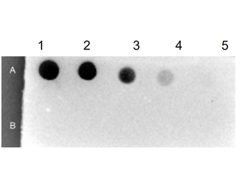
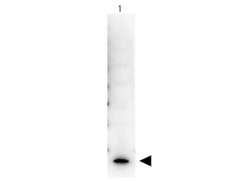
Dot Blot Results of Llama IgG2 Isotype control Biotin Conjugated. Llama IgG2 Isotype control Biotin Conjugate (1) 100 ng, (2) 33.33 ng, (3) 11.11 ng, (4) 3.70 ng, (5) 1.23. Antibody: Streptavidin (p/n orb348765) at 1:40000 for 30 mins at RT. Block: BlockOut (p/n orb348644) for 30 mins at RT. Exposure: 1 sec.
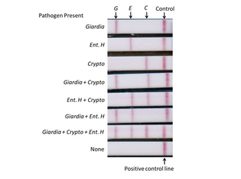
Multiplex lateral flow detection strips with three detection zones and a positive control zone. Strips tested positive(shown from top to bottom) for Giardia, Entamoeba histolytica, Cryptosporidium, Giardia + Cryptosporidium, Entamoeba histolytica + Cryptosporidium, Giardia + Entamoeba histolytica, and Giardia + Cryptosporidium + Entamoeba histolytica, and no pathogens. Streptavidin-coated gold colloid for lateral flow strips (Streptavidin p/n orb348765).
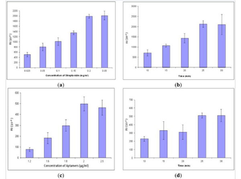
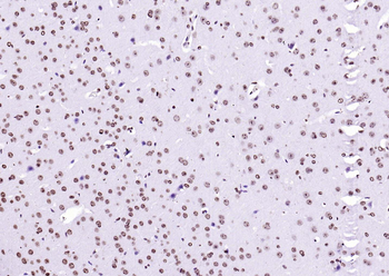
Optimization of streptavidin and aptamers, each concentration and incubation time repeated three times, respectively: (a) Streptavidin concentration; (b) Streptavidin incubation time; (c) Aptamer concentration; (d) Aptamer incubation time. Streptavidin (p/n orb348765).
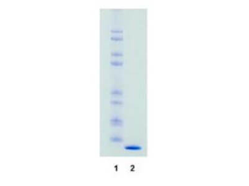
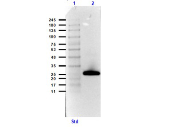
SDS-Page of Streptavidin. Lane 1: Molecular weight markers. Lane 2: Streptavidin. Load: 1.0 ug per lane. Predicted/Observed size: The molecular weight of streptavidin is 55000 daltons. The protein is composed of 4 essentially identical polypeptide chains (homotetramer). This product is chromatographically pure Streptavidin and shows predominantly a single 13.800 dalton band by SDS-PAGE.
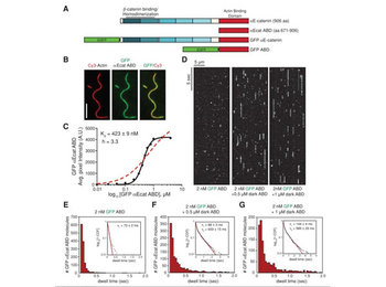
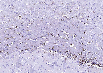
αE-catenin ABD binds cooperatively to actin filaments. (A) αE-catenin is composed of an array of five four-helix bundles (blue-shaded boxes) and a C-terminal five-helix bundle (red box). The β-catenin/homodimerization region and actin-binding domain are marked. All αE-catenin constructs used in this study are defined. (B) Localization of 1 µm GFP αE-catenin ABD bound to phalloidin-stabilized filamentous actin (20% Cy3 labeled). Scale bar, 5 µm. (C) Average fluorescence signal of GFP αE-catenin ABD bound to single-actin filaments plotted against total concentration of GFP αE-catenin ABD. Each data point represents average GFP fluorescence per pixel measured over ≥ 100 µm of single actin filaments (≥ 2 TIRF flow chambers). Data were fitted to either a Hill equation (black, straight line) or a hyperbolic function (red, dashed line). (D) Kymographs showing 2 nM GFP αE-catenin ABD binding and dissociating from the sides of single actin filaments in the absence or presence of 0.5 or 1 µm dark αE-catenin ABD. (E–G) Histograms of 2 nM GFP αE-catenin ABD dwell times on filamentous actin in the absence (E) or presence (F) of 0.5 µm dark αE-catenin ABD or (G) 1 µm dark αE-catenin ABD. Inset, curve fit of the 1-cumulative distribution frequency: (E) single-exponential fit (τ1 = 70 ± 2 ms, n = 1244 molecules), (F) double-exponential fit (τ1 = 88 ± 3 ms [58%)], τ2 = 659 ± 15 ms [42%], n = 1289 molecules), and (G) double-exponential fit (τ1 = 144 ± 4 ms [64%], τ2 = 986 ± 26 ms [36%], n = 1210 molecules). Streptavidin p/n orb348765).
Quick Database Links
UniProt
RefSeq:CAA00084.1
UniProt Details
− No UniProt data available
NCBI Reference Sequences
−Associated Accession Numbers
Curated reference sequences for the gene transcript and protein product| RefSeq | CAA00084.1 |
|---|
Documents Download
Datasheet
Product Information
Request a Document
Protocol Information
WB
Western Blot (IB, immunoblot)
ELISA
Enzyme-linked Immunosorbent Assay (EIA)
SDS-PAGE
Sodium Dodecyl Sulphate PolyAcrylamide Gel Electrophoresis
Streptavidin (orb348765)

- 0.0
Based on 0 reviews
Participating in our Biorbyt product reviews program enables you to support fellow scientists by sharing your firsthand experience with our products.
Login to Submit a ReviewAvailable Sizes
Select a size below