You have no items in your shopping cart.
Description
Research Area
Cancer Biology
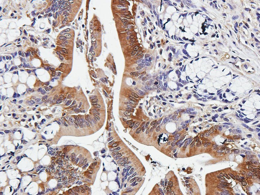
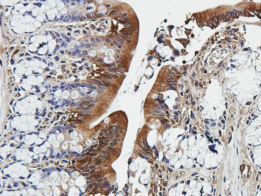
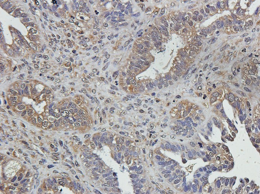
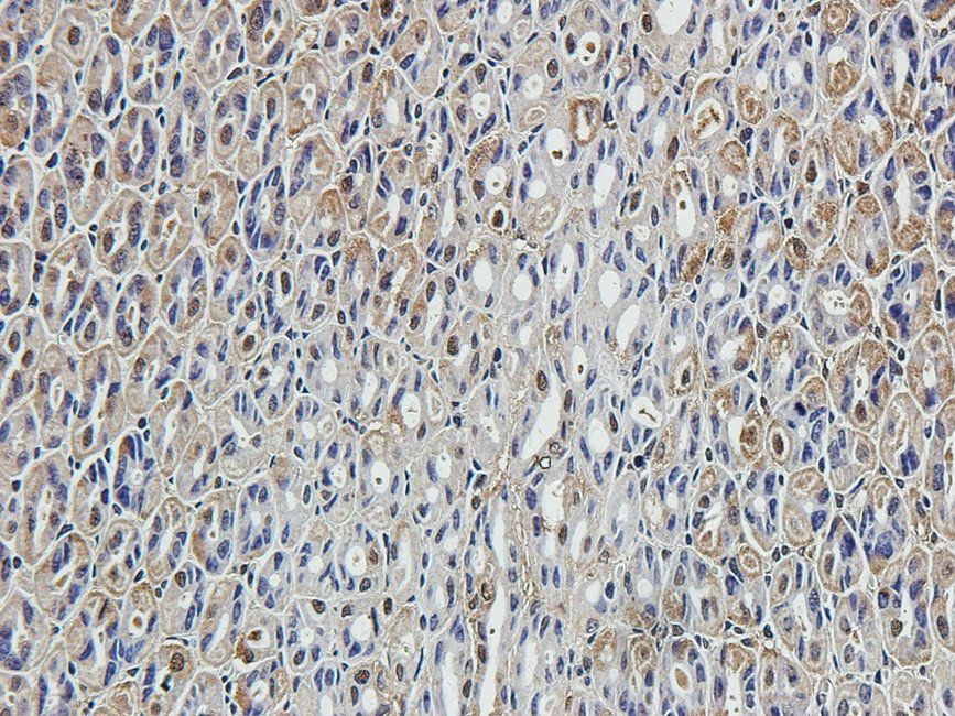
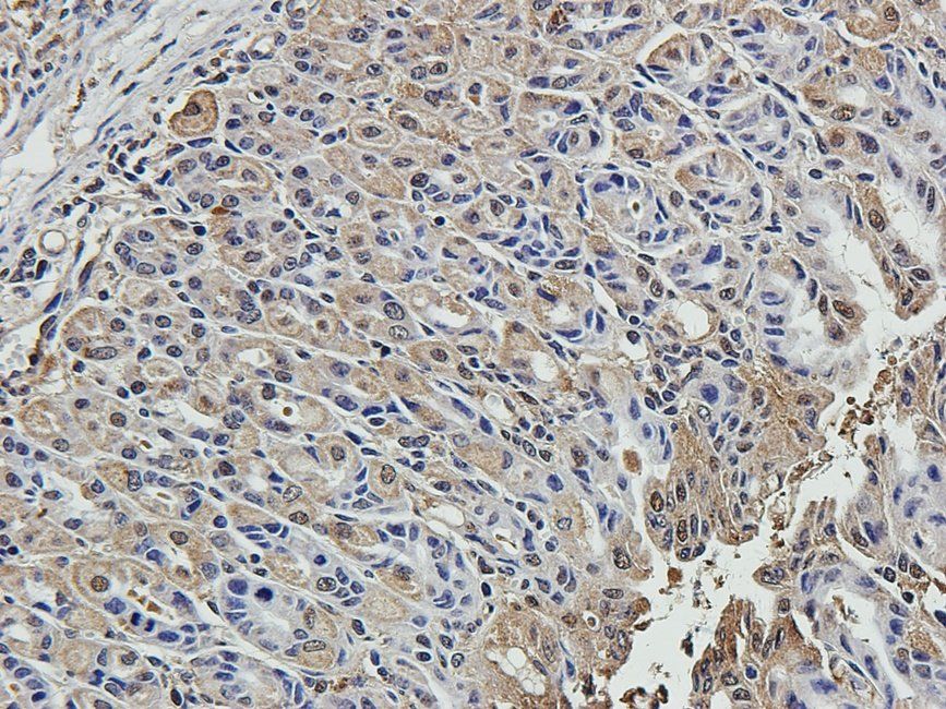
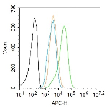
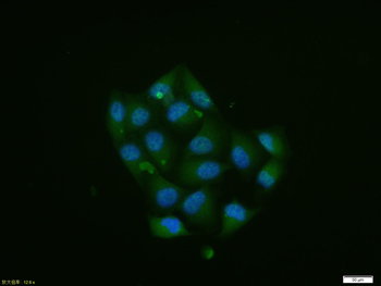
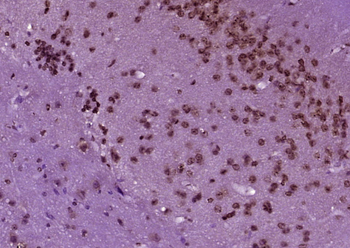
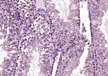
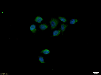
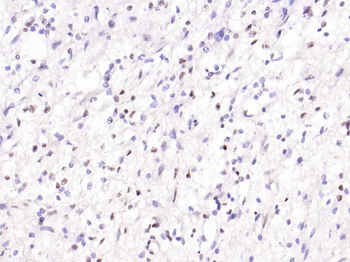
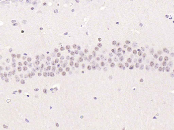
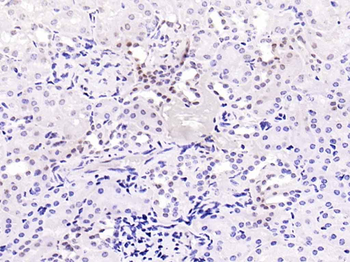
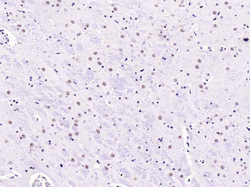
Images & Validation
−Item 1 of 1
| Tested Applications | ELISA, WB |
|---|---|
| Dilution Range | ELISA: 1:100,000, WB: 1:1,000 - 1:3,000 |
| Reactivity | Human, Monkey |
| Application Notes |
Key Properties
−| Antibody Type | Primary Antibody |
|---|---|
| Host | Rabbit |
| Clonality | Polyclonal |
| Isotype | IgG |
| Immunogen | This affinity purified antibody was prepared from whole rabbit serum produced by repeated immunizations with a synthetic peptide corresponding to an internal region of human Smad2 protein. |
| Target | SMAD2 |
| Purity | This affinity purified antibody is directed against human Smad2 protein. The product was affinity purified from monospecific antiserum by immunoaffinity chromatography. A BLAST analysis was used to suggest cross-reactivity with Smad2 protein from human, mouse and rat based on 100% homology with the immunizing sequence. Reactivity against homologues from other sources is not known. Also, the antibody is Smad2 specific, and reactivity to other Smad proteins (specifically Smad1, Smad3, Smad4, and Smad7) is not detected in over-expressed cell lysates (Personal Communication, Kathleen Flanders, CCR-NCI, Bethesda, MD). |
| Conjugation | Unconjugated |
Storage & Handling
−| Storage | Store vial at -20° C or below prior to opening. This vial contains a relatively low volume of reagent (25 µL). To minimize loss of volume dilute 1:10 by adding 225 µL of the buffer stated above directly to the vial. Recap, mix thoroughly and briefly centrifuge to collect the volume at the bottom of the vial. Use this intermediate dilution when calculating final dilutions as recommended below. Store the vial at -20°C or below after dilution. Avoid cycles of freezing and thawing. |
|---|---|
| Form/Appearance | Liquid (sterile filtered) |
| Buffer/Preservatives | Preservative: 0.01% (w/v) Sodium Azide. Stabilizer: None; Buffer: 0.02 M Potassium Phosphate, 0.15 M Sodium Chloride, pH 7.2 |
| Concentration | 0.55 mg/mL |
| Expiration Date | 12 months from date of receipt. |
| Dry Ice Shipping | Please note: This product requires shipment on dry ice. A dry ice surcharge will apply. |
| Disclaimer | For research use only |
Alternative Names
−rabbit anti-SMAD2 antibody, SMAD-2, SMAD 2, mothers against decapentaplegic homolog 2 antibody, MAD homolog 2, Mothers against DPP homolog 2, SMAD family member 2, MADH2, MADH 2, JV18-1
Similar Products
−- Item 1 of 10
SMAD2 Rabbit Polyclonal Antibody [orb507565]
IHC-P, WB
Guinea pig, Human, Mouse, Rat
Rabbit
Polyclonal
Unconjugated
100 μg - Item 1 of 5
Phospho-Smad2 (Ser465 + Ser467) Rabbit Polyclonal Antibody [orb6979]
FC, ICC, IF, IHC-Fr, IHC-P, WB
Bovine, Canine, Equine, Gallus, Porcine
Human, Mouse, Rat
Rabbit
Polyclonal
Unconjugated
50 μl, 100 μl, 200 μl - Item 1 of 8
Phospho-Smad2 (Ser467) Rabbit Polyclonal Antibody [orb783427]
IF, IHC-Fr, IHC-P
Mouse, Rat
Human, Mouse, Rat
Rabbit
Polyclonal
Unconjugated
50 μl, 100 μl, 200 μl - Item 1 of 5
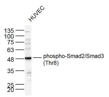
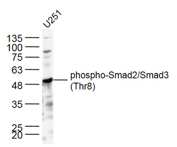
Phospho-Smad2/Smad3 (Thr8) Rabbit Polyclonal Antibody [orb186235]
FC, WB
Bovine, Canine, Equine, Gallus, Porcine, Rat
Human, Mouse
Rabbit
Polyclonal
Unconjugated
50 μl, 100 μl, 200 μl - Item 1 of 7

Quality Guarantee
Explore bioreagents carefree to elevate your research. All our products are rigorously tested for performance. If a product does not perform as described on its datasheet, our scientific support team will provide expert troubleshooting, a prompt replacement, or a refund. For full details, please see our Terms & Conditions and Buying Guide. Contact us at [email protected].
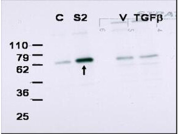
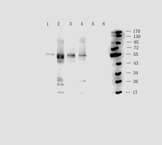
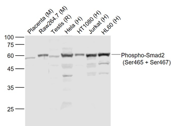
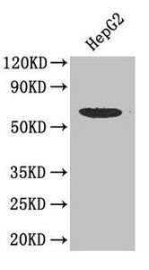
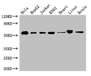
Western blot using Biorbyt's affinity purified anti-Smad2 to detect over-expressed Smad2 in COS cells (arrow). Lane C shows mock infection of COS cells with lentiviral vector alone. Lane S2 shows detection of Smad2 in lysates of COS transfected with Smad2. Lane V contains lysates of MDA-MB231 cells treated with vehicle; the next lane contains lysates of MDA-MB231 cells treated with TGF beta. Low levels of staining in control lanes correspond to detection of endogenous Smad2. Pre-incubation of the antibody with immunizing peptide (data not shown) completely blocks specific band staining. The blot presented is askew relative to the molecular weight markers. The expected MW for Smad2 is 52 kDa. The membrane was probed with the primary antibody at a 1:2500 dilution.
Documents Download
Datasheet
Product Information
Request a Document
Protocol Information
WB
Western Blot (IB, immunoblot)
ELISA
Enzyme-linked Immunosorbent Assay (EIA)
SMAD2 Antibody (orb345610)

- 0.0
Based on 0 reviews
Participating in our Biorbyt product reviews program enables you to support fellow scientists by sharing your firsthand experience with our products.
Login to Submit a ReviewAvailable Sizes
Select a size below
Free Secondary Antibody (20 ul)0/0
Please add an antibody product to your cart first.