You have no items in your shopping cart.
Featured
Description
Images & Validation
−Item 1 of 3
| Tested Applications | IF, IHC-P, WB |
|---|---|
| Dilution Range | IF - 1:10-50, WB - 1:1000, IHC-P - 1:10-50 |
| Reactivity | Human |
| Predicted Reactivity | Mouse |
Key Properties
−| Antibody Type | Primary Antibody |
|---|---|
| Host | Rabbit |
| Clonality | Polyclonal |
| Isotype | Rabbit IgG |
| Immunogen | Synthetic Peptide |
| Target | SIX5 |
| Molecular Weight | 74562 |
| Conjugation | Unconjugated |
Storage & Handling
−| Storage | Maintain refrigerated at 2-8°C for up to 2 weeks. For long term storage store at -20°C in small aliquots to prevent freeze-thaw cycles |
|---|---|
| Form/Appearance | Purified polyclonal antibody supplied in PBS with 0.09% (W/V) sodium azide. This antibody is purified through a protein A column, followed by peptide affinity purification. |
| Expiration Date | 12 months from date of receipt. |
| Disclaimer | For research use only |
Alternative Names
−Anti-Homeobox protein SIX5 antibody, anti-SIX5 antibody, anti-DMAHP antibody
Similar Products
−- Item 1 of 1
SIX5 Rabbit Polyclonal Antibody [orb665786]
WB
Human, Mouse
Rabbit
Polyclonal
Unconjugated
30 μl, 100 μl, 200 μl, 50 μl
Quality Guarantee
Explore bioreagents carefree to elevate your research. All our products are rigorously tested for performance. If a product does not perform as described on its datasheet, our scientific support team will provide expert troubleshooting, a prompt replacement, or a refund. For full details, please see our Terms & Conditions and Buying Guide. Contact us at [email protected].
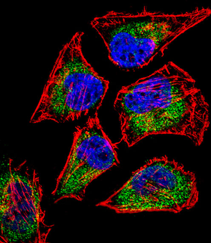
Fluorescent confocal image of Hela cell stained with SIX5 Antibody (Center). Hela cells were fixed with 4% PFA (20 min), permeabilized with Triton X-100 (0.1%, 10 min), then incubated with SIX5 primary antibody (1:25, 1 h at 37°C). For secondary antibody, Alexa Fluor 488 conjugated donkey anti-rabbit antibody (green) was used (1:400, 50 min at 37°C). Cytoplasmic actin was counterstained with Alexa Fluor 555 (red) conjugated Phalloidin (7 units/ml, 1 h at 37°C). Nuclei were counterstained with DAPI (blue) (10 µg/ml, 10 min). SIX5 immunoreactivity is localized to Cytoplasm significantly.
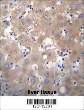
SIX5 Antibody (Center) immunohistochemistry analysis in formalin fixed and paraffin embedded human liver tissue followed by peroxidase conjugation of the secondary antibody and DAB staining. This data demonstrates the use of SIX5 Antibody (Center) for immunohistochemistry. Clinical relevance has not been evaluated.
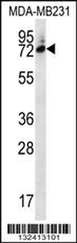
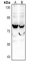
SIX5 Antibody (Center) western blot analysis in MDA-MB231 cell line lysates (35 ug/lane). This demonstrates the SIX5 antibody detected the SIX5 protein (arrow).
Quick Database Links
UniProt Details
− No UniProt data available
NCBI Reference Sequences
−Associated Accession Numbers
Curated reference sequences for the gene transcript and protein product| Protein | NP_787071.2 |
|---|
Documents Download
Datasheet
Product Information
Request a Document
Protocol Information
WB
Western Blot (IB, immunoblot)
IHC-P
Immunohistochemistry Paraffin
IF
Immunofluorescence
Filter by Applications
Filter by Species
Dan Zhou 1, Yuqing Song 2, Liang Chang Role of SIX5-mediated EXO1 overexpression in driving glioblastoma progression: Insights into tumor cell migration and angiogenesis Brain Res, (2025)
Applications
WB
Reactivity
Human
SIX5 Antibody (Center) (orb30764)

- 0.0
Based on 0 reviews
Participating in our Biorbyt product reviews program enables you to support fellow scientists by sharing your firsthand experience with our products.
Login to Submit a ReviewAvailable Sizes
Select a size below
Free Secondary Antibody (20 ul)0/0
Please add an antibody product to your cart first.