You have no items in your shopping cart.
Featured
Description
Research Area
Neuroscience
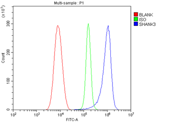
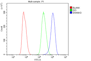
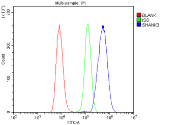
Images & Validation
−Item 1 of 4
| Tested Applications | AM, ICC, IF, IHC, IP, WB |
|---|---|
| Dilution Range | WB (1:1000), IHC-P (1:100), ICC/IF (1:100) |
| Reactivity | Human, Mouse, Rat |
| Application Notes |
Key Properties
−| Antibody Type | Recombinant Antibody |
|---|---|
| Host | Mouse |
| Clonality | Recombinant |
| Isotype | IgG2b |
| Clone No. | S69 |
| Immunogen | Synthetic peptide amino acids 840-857 of rat Shank3 |
| Target | SHANK3 |
| Molecular Weight | 190kDa |
| Purification | Protein G Purified |
| Conjugation | Unconjugated |
Storage & Handling
−| Storage | Maintain refrigerated at 2-8°C for up to 2 weeks. For long term storage store at -20°C in small aliquots to prevent freeze-thaw cycles. |
|---|---|
| Buffer/Preservatives | PBS pH 7.4, 50% glycerol, 0.09% sodium azide. Storage buffer changes when conjugated. |
| Concentration | 1 mg/ml |
| Expiration Date | 12 months from date of receipt. |
| Disclaimer | For research use only |
Alternative Names
−AI841104, DEL22q13.3, KIAA1650, Proline rich synapse associated protein 2, Proline-rich synapse-associated protein 2, ProSAP2, PSAP2, SH3 and multiple ankyrin repeat domains 3, SH3 and multiple ankyrin repeat domains protein 3, SH3/ankyrin domain gene 3, SHAN3_HUMAN, Shank postsynaptic density protein, Shank3, Shank3b, SPANK 2, SPANK2
Similar Products
−- Item 1 of 6
SHANK3 Rabbit Polyclonal Antibody [orb669123]
ELISA, FC, IHC, WB
Human, Mouse, Rat
Rabbit
Polyclonal
Unconjugated
100 μg - Item 1 of 4
SHANK3 Antibody (APC) [orb148738]
AM, ICC, IF, IHC, IP, WB
Human, Mouse, Rat
Mouse
Recombinant
APC
100 μg - Item 1 of 4
SHANK3 Antibody (Biotin) [orb148739]
AM, ICC, IF, IHC, IP, WB
Human, Mouse, Rat
Mouse
Recombinant
Biotin
100 μg - Item 1 of 4
SHANK3 Antibody (FITC) [orb148740]
AM, ICC, IF, IHC, IP, WB
Human, Mouse, Rat
Mouse
Recombinant
FITC
100 μg - Item 1 of 4
SHANK3 Antibody (HRP) [orb148741]
AM, ICC, IF, IHC, IP, WB
Human, Mouse, Rat
Mouse
Recombinant
HRP
100 μg


Quality Guarantee
Explore bioreagents carefree to elevate your research. All our products are rigorously tested for performance. If a product does not perform as described on its datasheet, our scientific support team will provide expert troubleshooting, a prompt replacement, or a refund. For full details, please see our Terms & Conditions and Buying Guide. Contact us at [email protected].

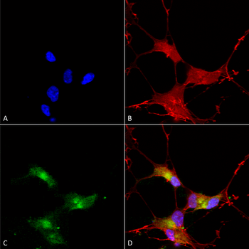
Immunocytochemistry/Immunofluorescence analysis using Mouse Anti-SHANK3 Monoclonal Antibody, Clone S69-46. Tissue: Neuroblastoma cells (SH-SY5Y). Species: Human. Fixation: 4% PFA for 15 min. Primary Antibody: Mouse Anti-SHANK3 Monoclonal Antibody at 1:50 for overnight at 4°C with slow rocking. Secondary Antibody: AlexaFluor 488 at 1:1000 for 1 hour at RT. Counterstain: Phalloidin-iFluor 647 (red) F-Actin stain; Hoechst (blue) nuclear stain at 1:800, 1.6mM for 20 min at RT. (A) Hoechst (blue) nuclear stain. (B) Phalloidin-iFluor 647 (red) F-Actin stain. (C) SHANK3 Antibody (D) Composite.
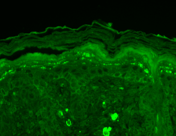
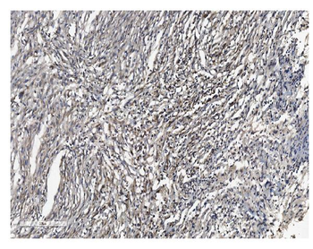
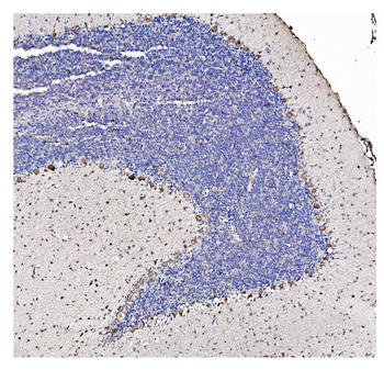
Immunohistochemistry analysis using Mouse Anti-SHANK3 Monoclonal Antibody, Clone S69-46. Tissue: backskin. Species: Mouse. Fixation: Bouin's Fixative and paraffin-embedded. Primary Antibody: Mouse Anti-SHANK3 Monoclonal Antibody at 1:100 for 1 hour at RT. Secondary Antibody: FITC Goat Anti-Mouse (green) at 1:50 for 1 hour at RT. Localization: Early stages of filaggrin-like and dermal staining.

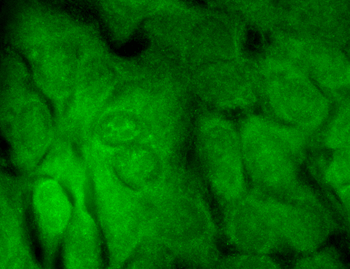
Immunocytochemistry/Immunofluorescence analysis using Mouse Anti-SHANK3 Monoclonal Antibody, Clone S69-46. Tissue: HaCaT cells. Species: Human. Fixation: Cold 100% methanol for 10 minutes at -20°C. Primary Antibody: Mouse Anti-SHANK3 Monoclonal Antibody at 1:100 for 1 hour at RT. Secondary Antibody: FITC Goat Anti-Mouse (green) at 1:50 for 1 hour at RT. Localization: Borderline positive.

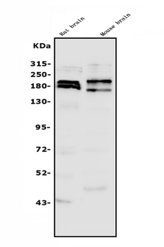
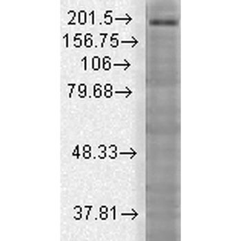
Western Blot analysis of Rat brain membrane lysate showing detection of SHANK3 protein using Mouse Anti-SHANK3 Monoclonal Antibody, Clone S69-46. Load: 15 μg. Block: 1.5% BSA for 30 minutes at RT. Primary Antibody: Mouse Anti-SHANK3 Monoclonal Antibody at 1:1000 for 2 hours at RT. Secondary Antibody: Sheep Anti-Mouse IgG: HRP for 1 hour at RT.
Quick Database Links
UniProt Details
− No UniProt data available
NCBI Gene Details
− No NCBI Gene data available
NCBI Reference Sequences
−Associated Accession Numbers
Curated reference sequences for the gene transcript and protein product| Protein | NP_067708.1 |
|---|
Documents Download
Datasheet
Product Information
Request a Document
Protocol Information
WB
Western Blot (IB, immunoblot)
IHC
Immunohistochemistry
IF
Immunofluorescence
ICC
Immunocytochemistry
IP
Immunoprecipitation
SHANK3 Antibody (orb67424)

- 0.0
Based on 0 reviews
Participating in our Biorbyt product reviews program enables you to support fellow scientists by sharing your firsthand experience with our products.
Login to Submit a ReviewAvailable Sizes
Select a size below
Choose Conjugation or Carrier Free Version
Free Secondary Antibody (20 ul)0/0
Please add an antibody product to your cart first.