You have no items in your shopping cart.
Featured
Description
Research Area
Neuroscience
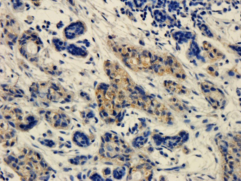
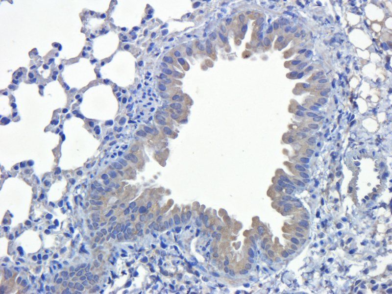
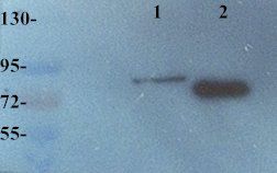
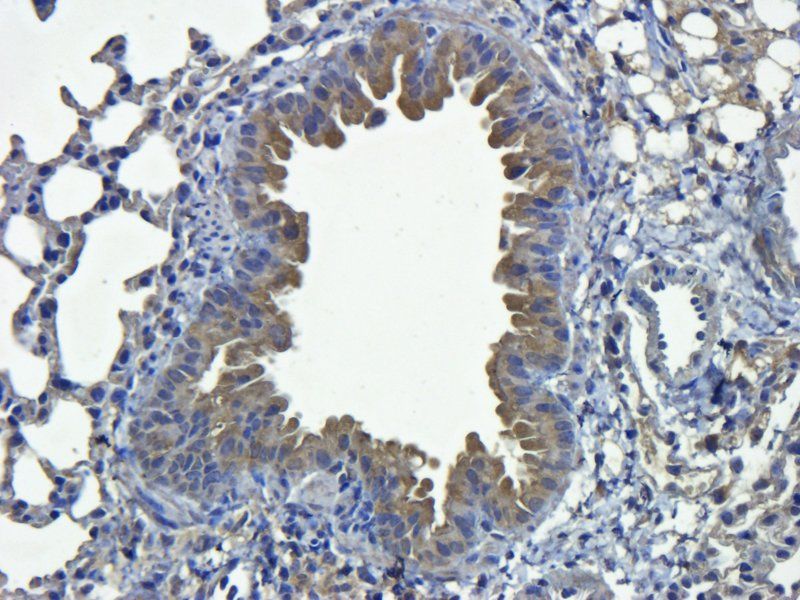
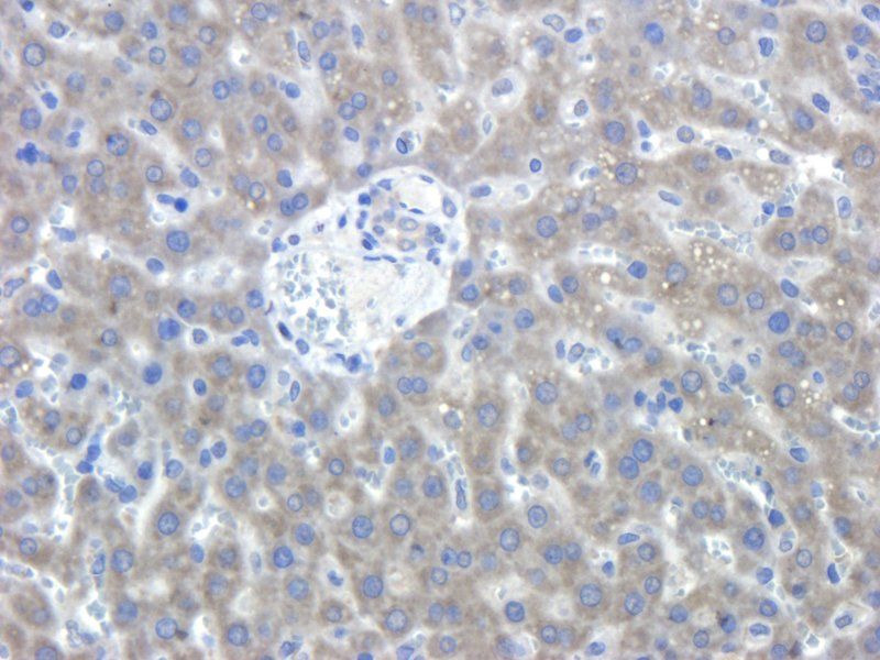
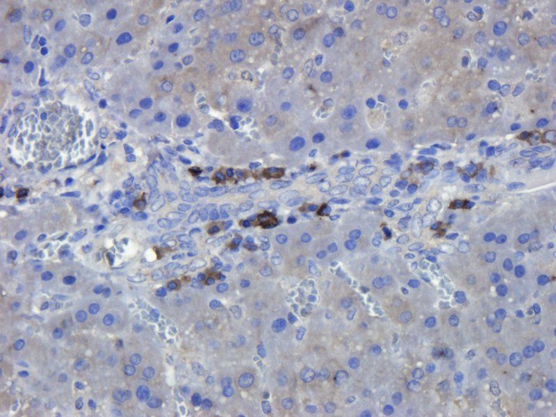
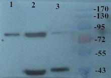
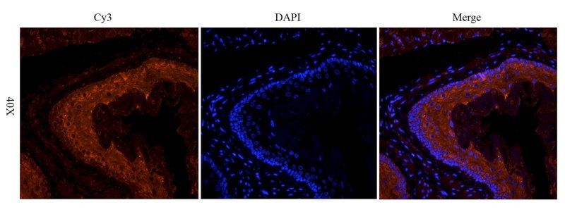
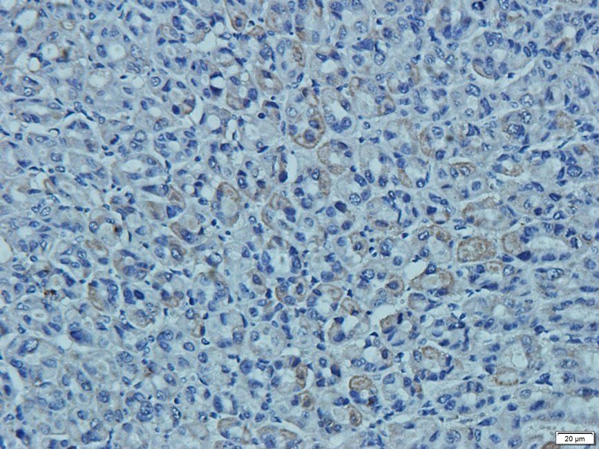
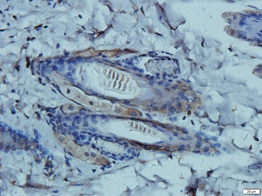
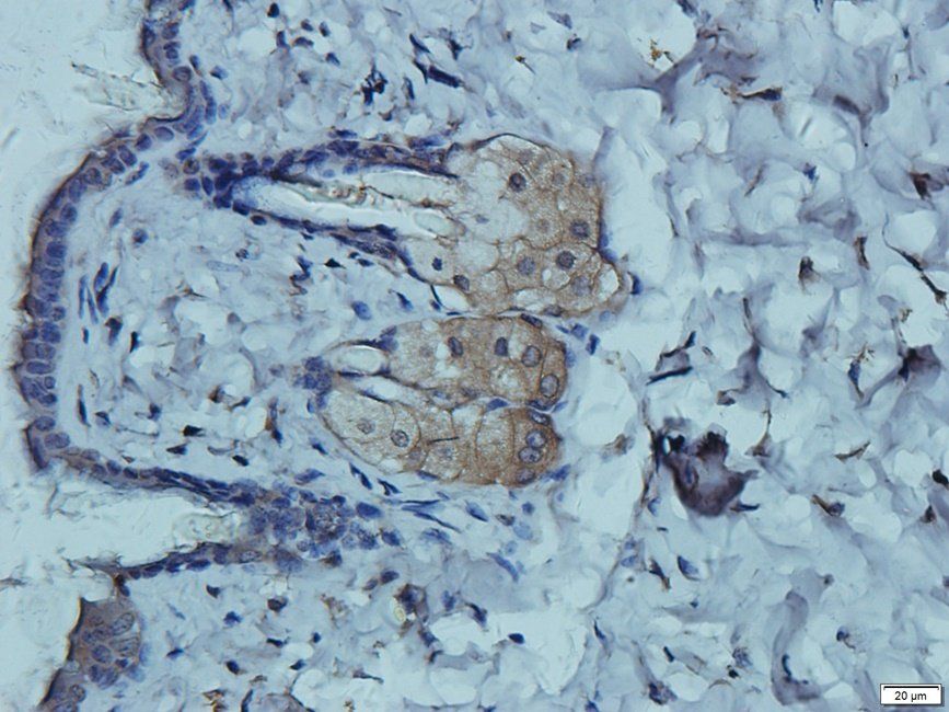
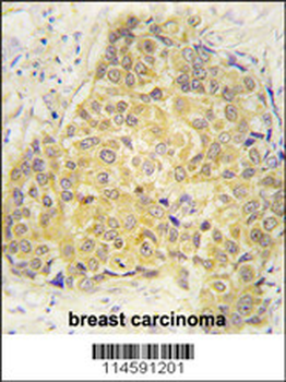
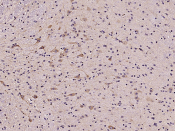
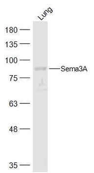
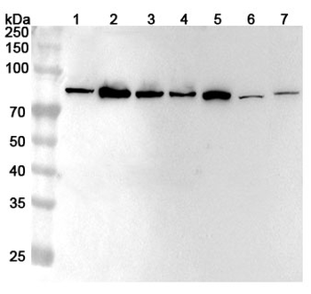
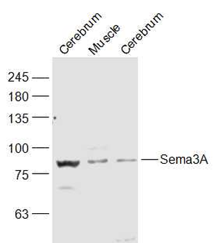
Images & Validation
−

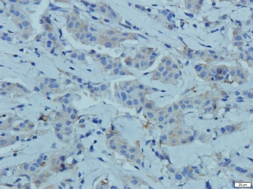
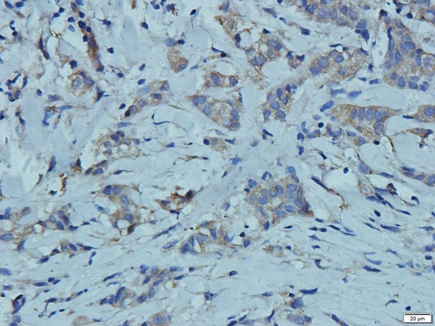
Item 1 of 15
| Tested Applications | ICC, IF, IHC-P, WB |
|---|---|
| Dilution Range | WB: 1:100-800, IHC-P: 1: 100-500 |
| Reactivity | Human, Mouse, Rat |
Key Properties
−| Host | Rabbit |
|---|---|
| Clonality | Polyclonal |
| Isotype | IgG |
| Immunogen | KLH conjugated synthetic peptide derived from human SEMA3A. Please contact us for the exact immunogen sequence. The peptide is available as orb374747. |
| Target | SEMA3A |
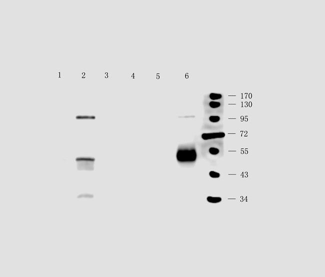
| Molecular Weight | 89 kDa |
| Purity | Polyclonal antibodies are purified by peptide affinity chromatography |
| Conjugation | Unconjugated |
Storage & Handling
−| Storage | Maintain refrigerated at 2-8°C for up to 2 weeks. For long term storage store at -20°C in small aliquots to prevent freeze-thaw cycles. |
|---|---|
| Form/Appearance | 10 mM PBS, 0.02% sodium azide |
| Concentration | - 100 μg (in 200 μl): 0.5 mg/ml- 200 μg (in 400 μl): 0.5 mg/ml |
| Expiration Date | 12 months from date of receipt. |
| Disclaimer | For research use only |
Alternative Names
−anti Coll 1 antibody, anti Coll1 antibody, anti HH16 antibody, anti Hsema I antibody, anti Hsema III antibody, anti Sema III antibody, anti SEMA1 antibody, anti SEMA1-PEN antibody, anti SEMA3A antibody, anti SEMAD antibody, anti SEMAIII antibody, anti SEMAL antibody, anti Semaphorin 3A precursor antibody, anti Semaphorin III antibody, anti Semaphorin-3A precursor antibody, anti Semaphorin3A antibody, anti SemD antibody
Similar Products
−- Item 1 of 2
SEMA3A Antibody (Center) [orb1928916]
IHC-P, WB
Mouse, Rat
Human
Rabbit
Polyclonal
Unconjugated
50 μl, 100 μl - Item 1 of 2
Sema3A Rabbit Polyclonal Antibody [orb313757]
WB
Canine, Equine, Gallus, Human, Porcine, Rabbit, Rat, Sheep
Mouse
Rabbit
Polyclonal
Unconjugated
50 μl, 100 μl, 200 μl - Item 1 of 1
SEMA3A Rabbit Polyclonal Antibody [orb2950817]
ELISA, IHC, WB
Human
Rabbit
Polyclonal
Unconjugated
50 μg, 100 μg - Item 1 of 2
- Item 1 of 1
Sema3A Rabbit Polyclonal Antibody [orb186022]
WB
Canine, Equine, Gallus, Human, Porcine, Rabbit, Sheep
Mouse, Rat
Rabbit
Polyclonal
Unconjugated
50 μl, 100 μl, 200 μl

Quality Guarantee
Explore bioreagents carefree to elevate your research. All our products are rigorously tested for performance. If a product does not perform as described on its datasheet, our scientific support team will provide expert troubleshooting, a prompt replacement, or a refund. For full details, please see our Terms & Conditions and Buying Guide. Contact us at [email protected].
Quick Database Links
Protocol Information
WB
Western Blot (IB, immunoblot)
IHC-P
Immunohistochemistry Paraffin
IF
Immunofluorescence
ICC
Immunocytochemistry
Available Sizes
Select a size below
Choose Conjugation or Carrier Free Version
Free Secondary Antibody (20 ul)0/0
Please add an antibody product to your cart first.