You have no items in your shopping cart.
Description
Research Area
Infectious Disease & Virology
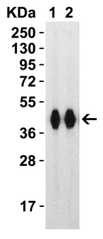
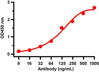
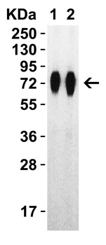
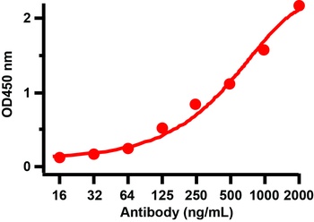
Images & Validation
−
Item 1 of 14
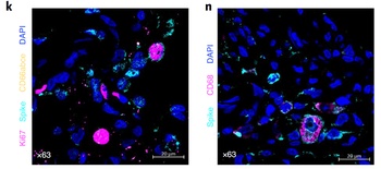
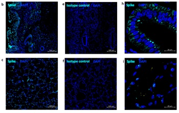
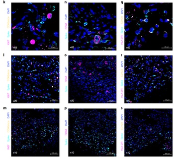
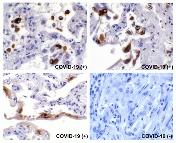
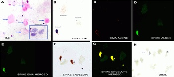
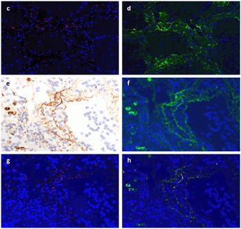
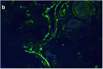
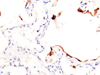
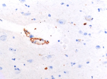
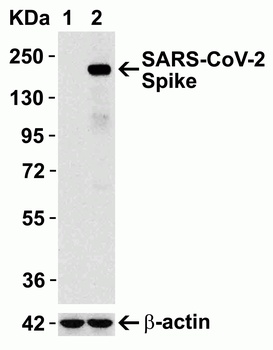
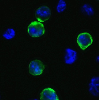
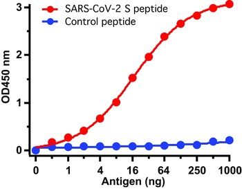
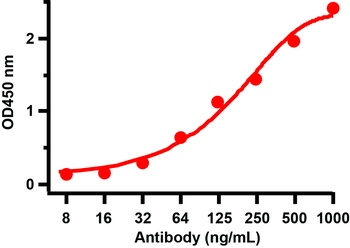
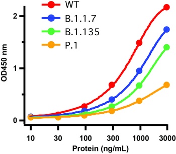
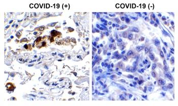
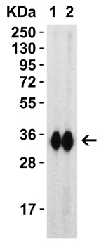
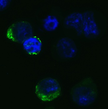
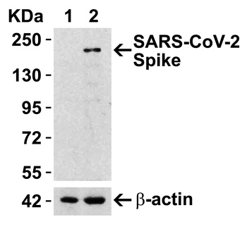
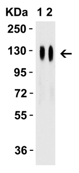
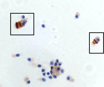
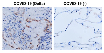
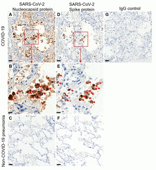
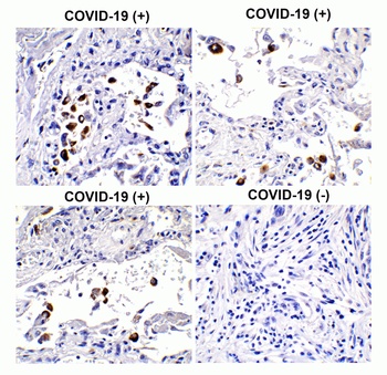
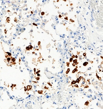
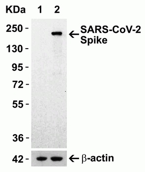
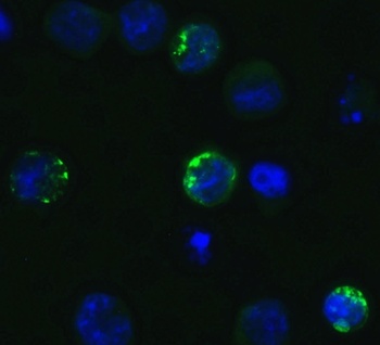
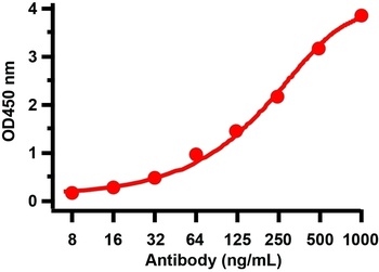
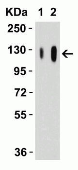
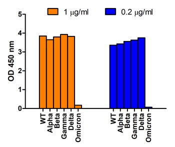
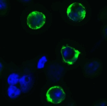
| Tested Applications | ELISA, IF, IHC, WB |
|---|---|
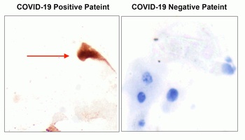
| Reactivity | Virus |
| Application Notes |
Key Properties
−| Antibody Type | Primary Antibody |
|---|---|
| Host | Rabbit |
| Clonality | Polyclonal |
| Isotype | IgG |
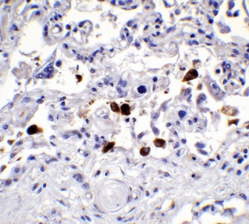
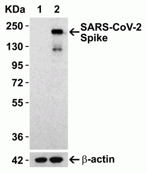
| Immunogen | Anti-SARS-CoV-2 (COVID-19) Spike antibody (orb1239976) was raised against a peptide corresponding to 20 amino acids near the carboxy terminus of SARS-CoV-2 (COVID-19) Spike glycoprotein. The immunogen is located within the last 50 amino acids of SARS-CoV-2 (COVID-19) Spike protein. |
| Target | S |
| Purification | SARS-CoV-2 (COVID-19) Spike Antibody is affinity chromatography purified via peptide column. |
| Conjugation | Unconjugated |
Storage & Handling
−| Storage | Maintain refrigerated at 2-8°C for up to 2 weeks. For long term storage store at -20°C in small aliquots to prevent freeze-thaw cycles. |
|---|---|
| Form/Appearance | Liquid |
| Buffer/Preservatives | SARS-CoV-2 (COVID-19) Spike Antibody is supplied in PBS containing 0.02% sodium azide. |
| Concentration | 1 mg/mL |
| Expiration Date | 12 months from date of receipt. |
| Disclaimer | For research use only |
Alternative Names
−SARS-CoV-2 (COVID-19) Spike Antibody: Severe acute respiratory syndrome coronavirus 2 (SARS-CoV-2), Surface Glycoprotein, Spike protein
Similar Products
−- Item 1 of 11
SARS-CoV-2 (COVID-19) Spike RBD Antibody [orb1239994]
ELISA, IF, IHC, WB
Virus
Rabbit
Polyclonal
Unconjugated
0.02 mg, 0.1 mg - Item 1 of 9
SARS-CoV-2 (COVID-19) Spike S1 Antibody [orb1239995]
ELISA, ICC, IF, IHC, WB
Virus
Rabbit
Polyclonal
Unconjugated
0.02 mg, 0.1 mg - Item 1 of 8
SARS-CoV-2 (COVID-19) Spike 681P Antibody [orb1239981]
ELISA, IF, WB
Virus
Rabbit
Polyclonal
Unconjugated
0.1 mg, 0.02 mg - Item 1 of 4
Anti-COVID-19 & SARS-CoV S glycoprotein [CR3022] [orb758974]
ELISA, IF
Virus
Human
Monoclonal
Unconjugated
0.05 mg - Item 1 of 4
Anti-COVID-19 & SARS-CoV S glycoprotein [CR3022] [orb758976]
ELISA, IF
Virus
Human
Monoclonal
Unconjugated
0.2 mg

![Anti-COVID-19 & SARS-CoV S glycoprotein [CR3022]](/images/pub/media/catalog/product/NewWebsite/35/orb758974_1.png)
![Anti-COVID-19 & SARS-CoV S glycoprotein [CR3022]](/images/pub/media/catalog/product/NewWebsite/35/orb758974_2.png)
![Anti-COVID-19 & SARS-CoV S glycoprotein [CR3022]](/images/pub/media/catalog/product/NewWebsite/35/orb758974_3.png)
![Anti-COVID-19 & SARS-CoV S glycoprotein [CR3022]](/images/pub/media/catalog/product/NewWebsite/35/orb758974_4.png)
![Anti-COVID-19 & SARS-CoV S glycoprotein [CR3022]](/images/pub/media/catalog/product/NewWebsite/35/orb758976_1.png)
![Anti-COVID-19 & SARS-CoV S glycoprotein [CR3022]](/images/pub/media/catalog/product/NewWebsite/35/orb758976_2.png)
![Anti-COVID-19 & SARS-CoV S glycoprotein [CR3022]](/images/pub/media/catalog/product/NewWebsite/35/orb758976_3.png)
![Anti-COVID-19 & SARS-CoV S glycoprotein [CR3022]](/images/pub/media/catalog/product/NewWebsite/35/orb758976_4.png)
Quality Guarantee
Explore bioreagents carefree to elevate your research. All our products are rigorously tested for performance. If a product does not perform as described on its datasheet, our scientific support team will provide expert troubleshooting, a prompt replacement, or a refund. For full details, please see our Terms & Conditions and Buying Guide. Contact us at [email protected].
Protocol Information
WB
Western Blot (IB, immunoblot)
IHC
Immunohistochemistry
IF
Immunofluorescence
ELISA
Enzyme-linked Immunosorbent Assay (EIA)
Available Sizes
Select a size below
Free Secondary Antibody (20 ul)0/0
Please add an antibody product to your cart first.