You have no items in your shopping cart.
Description
Images & Validation
−Item 1 of 12
| Tested Applications | ELISA, WB |
|---|---|
| Dilution Range | ELISA: 1:75,000 - 1:150,000, WB: 1:1,000 - 1:10,000 |
| Reactivity | Other |
| Application Notes |
Key Properties
−| Antibody Type | Primary Antibody |
|---|---|
| Host | Mouse |
| Clonality | Monoclonal |
| Isotype | IgG2a |
| Clone No. | 8E5.G7 |
| Immunogen | The immunogen is a Red Fluorescent Protein (RFP) fusion protein corresponding to the full-length amino acid sequence (234aa) derived from the mushroom anemone Discosoma. |
| Target | DsRed |
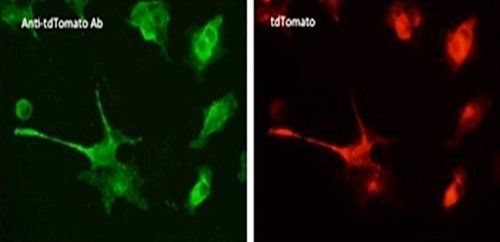
| Purity | Anti-RFP Monoclonal Antibody was purified from concentrated tissue culture supernate by Protein A chromatography. Expect reactivity against RFP and its variants: mCherry, tdTomato, mBanana, mOrange, mPlum, mOrange and mStrawberry. |
| Conjugation | Unconjugated |
Storage & Handling
−| Storage | Store Anti-RFP at -20° C or below prior to opening. This vial contains a relatively low volume of reagent (25 µL). To minimize loss of volume dilute 1:10 by adding 225 µL of the buffer stated above directly to the vial. Recap, mix thoroughly and briefly centrifuge to collect the volume at the bottom of the vial. Use this intermediate dilution when calculating final dilutions as recommended below. Store the vial at -20°C or below after dilution. Avoid cycles of freezing and thawing. |
|---|---|
| Form/Appearance | Liquid (sterile filtered) |
| Buffer/Preservatives | Preservative: 0.01% (w/v) Sodium Azide. Stabilizer: None; Buffer: 0.02 M Potassium Phosphate, 0.15 M Sodium Chloride, pH 7.2 |
| Concentration | 1.004 |
| Expiration Date | 12 months from date of receipt. |
| Dry Ice Shipping | Please note: This product requires shipment on dry ice. A dry ice surcharge will apply. |
| Disclaimer | For research use only |
Alternative Names
−mouse anti-RFP Antibody, DsRed, rDsRed, Discosoma sp. Red Fluorescent Protein, Red fluorescent protein drFP583
Similar Products
−- Item 1 of 45
DsRed Antibody [orb345391]
ELISA, FC, IF, IHC, KO/KD Validated, WB
Other
Rabbit
Polyclonal
Unconjugated
100 μg - Item 1 of 45
DsRed Antibody [orb345392]
ELISA, FC, IF, IHC, KO/KD Validated, WB
Other
Rabbit
Polyclonal
Unconjugated
25 μl - Item 1 of 2
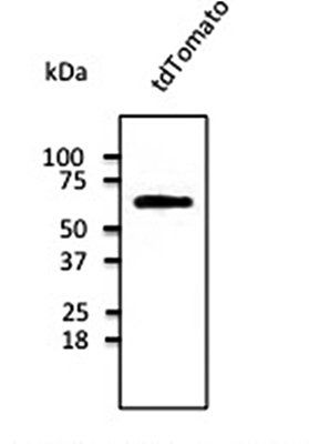
tdTomato Antibody [orb182397]
ELISA, FACS, IF, IHC-Fr, IHC-P, WB
Other
Goat
Polyclonal
Unconjugated
100 μg - Item 1 of 4
mCherry Goat Polyclonal Antibody [orb11618]
FC, ICC, IEM, IF, IHC-Fr, IHC-P, WB
Other
Goat
Polyclonal
Unconjugated
20 μg, 100 μg - Item 1 of 12


Quality Guarantee
Explore bioreagents carefree to elevate your research. All our products are rigorously tested for performance. If a product does not perform as described on its datasheet, our scientific support team will provide expert troubleshooting, a prompt replacement, or a refund. For full details, please see our Terms & Conditions and Buying Guide. Contact us at [email protected].

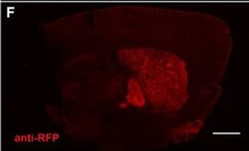
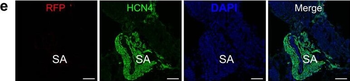
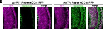
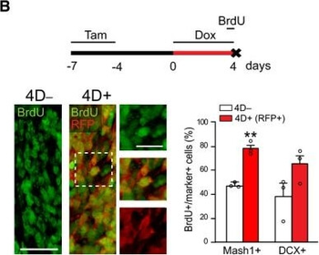
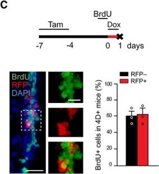
Characterization of the transgenic model and effect of 4D on the RMSA Fluorescence image of a sagittal section of a 4D+ brain after a 4‐day treatment with doxycycline showing RFP signal confined to the SVZ and RMS (nuclei counterstained with DAPI; blue). Insets show representative images of specific brain regions (as indicated) and the olfactory epithelium. A'Phase contrast picture of the SVZ upon in situ hybridization against mRNA for RFP in a 4D+ brain treated as in (A) and sacrificed immediately after (left) or 2 days after (right) doxycycline administration. (B, C) Experimental design (top), fluorescence pictures (left with magnified insets), and quantifications (right) of BrdU incorporation in the RMS (B) or SVZ (C). (B) shows the proportion of BrdU in C (Mash1+) and A (DCX+) cells in 4D− (white) and 4D+ (red; among RFP+) mice. (C) shows the proportion of RFP− (black) and RFP+ (red) among BrdU+ cells of 4D+ mice. (A) OB, olfactory bulb; RMS, rostral migratory stream; LV, lateral ventricle; DG, dentate gyrus; OE, olfactory epithelium. (A–C) Tam, tamoxifen; Dox, doxycycline. (B, C) Mean ± SEM; **P 1100 cells. Scale bars = 500 (A top, A'), 100 (insets A and A'), 50 (B and C), and 20 (insets B and C) µm.
Characterization of the transgenic model and effect of 4D on the RMSA Fluorescence image of a sagittal section of a 4D+ brain after a 4‐day treatment with doxycycline showing RFP signal confined to the SVZ and RMS (nuclei counterstained with DAPI; blue). Insets show representative images of specific brain regions (as indicated) and the olfactory epithelium. A'Phase contrast picture of the SVZ upon in situ hybridization against mRNA for RFP in a 4D+ brain treated as in (A) and sacrificed immediately after (left) or 2 days after (right) doxycycline administration. (B, C) Experimental design (top), fluorescence pictures (left with magnified insets), and quantifications (right) of BrdU incorporation in the RMS (B) or SVZ (C). (B) shows the proportion of BrdU in C (Mash1+) and A (DCX+) cells in 4D− (white) and 4D+ (red; among RFP+) mice. (C) shows the proportion of RFP− (black) and RFP+ (red) among BrdU+ cells of 4D+ mice. (A) OB, olfactory bulb; RMS, rostral migratory stream; LV, lateral ventricle; DG, dentate gyrus; OE, olfactory epithelium. (A–C) Tam, tamoxifen; Dox, doxycycline. (B, C) Mean ± SEM; **P 1100 cells. Scale bars = 500 (A top, A'), 100 (insets A and A'), 50 (B and C), and 20 (insets B and C) µm.
Characterization of the transgenic model and effect of 4D on the RMSA Fluorescence image of a sagittal section of a 4D+ brain after a 4‐day treatment with doxycycline showing RFP signal confined to the SVZ and RMS (nuclei counterstained with DAPI; blue). Insets show representative images of specific brain regions (as indicated) and the olfactory epithelium. A'Phase contrast picture of the SVZ upon in situ hybridization against mRNA for RFP in a 4D+ brain treated as in (A) and sacrificed immediately after (left) or 2 days after (right) doxycycline administration. (B, C) Experimental design (top), fluorescence pictures (left with magnified insets), and quantifications (right) of BrdU incorporation in the RMS (B) or SVZ (C). (B) shows the proportion of BrdU in C (Mash1+) and A (DCX+) cells in 4D− (white) and 4D+ (red; among RFP+) mice. (C) shows the proportion of RFP− (black) and RFP+ (red) among BrdU+ cells of 4D+ mice. (A) OB, olfactory bulb; RMS, rostral migratory stream; LV, lateral ventricle; DG, dentate gyrus; OE, olfactory epithelium. (A–C) Tam, tamoxifen; Dox, doxycycline. (B, C) Mean ± SEM; **P 1100 cells. Scale bars = 500 (A top, A'), 100 (insets A and A'), 50 (B and C), and 20 (insets B and C) µm.

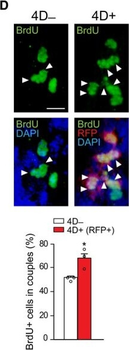
Chronic effect of 4D overexpression on NSC and OB neurogenesisAExperimental design used to assess the chronic effect of a transient 4D induction with BrdU and EdU given during Dox administration or 1 h before sacrifice, respectively.B–EFrom top to bottom: fluorescence pictures of the SVZ (B–D) or OB (E) and absolute number (B, C, and E) or proportions (C–E) of cells in 4D− (white bars) or 4D+ (black or red bars for all or RFP+ cells, respectively) mice scored positive for various markers as indicated. Insets in (C) are magnified (right) with arrowheads pointing label‐retaining NSC (white) or astrocytes (empty). Arrowheads in (D) point cell doublets (among RFP+ protein‐retaining cells in 4D+). (E) GL, glomerular; EPL, external plexiform; MCL, mitral cell and GCL, granule cell layers.Data information: (B–E) Mean ± SEM; *P 285 cells for each quantification. Scale bars = 50 µm (B, C, and E) and 20 µm (D and insets in C).

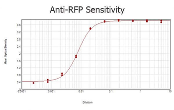
ELISA results of purified Mouse anti-RFP Monoclonal Antibody tested against RFP (p/n orb345960). Each well was coated in duplicate with 1.0 µg of the antigen. The starting dilution of antibody was 5 µg/ml and the X-axis represents the Log10 of a 3-fold dilution. This titration is a 4-parameter curve fit where the IC50 is defined as the titer of the antibody. Assay performed using 3% fish gel, anti-Mouse IgG Antibody Peroxidase Conjugated Secondary and TMB ELISA Peroxidase Substrate (p/n orb348651).

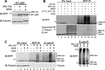
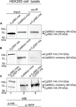
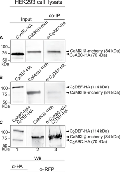
Immunoprecipitation and western blot show interaction of otoferlin with CaMKIIδ. (A–C) Two HA-tagged mouse otoferlin fragments, C2ABC (aa 1–632 in NP_001093865; 70 kDa) and C2DEF (aa 933–1920; 114 kDa) were co-transfected with mcherry-tagged mouse CaMKIIδ into HEK293 cells. Transfections were performed either with otoferlin C2ABC and CaMKIIδ (A, Input Lane 1 and 2), otoferlin C2DEF and CaMKIIδ (B, Input Lane 1 and 2) or in the presence of both C2ABC and C2DEF fragments and CaMKIIδ (C, Input Lane 1 and 2). Co-immunoprecipitations of C2ABC-HA and C2DEF-HA were conducted from HEK293 cell lysates using anti-HA antibodies. CaMKIIδ-mcherry was detected in the eluate using an anti-RFP (red fluorescent protein) antibody (A–C, Lane 3), indicating that CaMKIIδ co-precipitated with recombinant otoferlin fragments.
Immunoprecipitation and western blot show interaction of otoferlin with CaMKIIδ. (A–C) Two HA-tagged mouse otoferlin fragments, C2ABC (aa 1–632 in NP_001093865; 70 kDa) and C2DEF (aa 933–1920; 114 kDa) were co-transfected with mcherry-tagged mouse CaMKIIδ into HEK293 cells. Transfections were performed either with otoferlin C2ABC and CaMKIIδ (A, Input Lane 1 and 2), otoferlin C2DEF and CaMKIIδ (B, Input Lane 1 and 2) or in the presence of both C2ABC and C2DEF fragments and CaMKIIδ (C, Input Lane 1 and 2). Co-immunoprecipitations of C2ABC-HA and C2DEF-HA were conducted from HEK293 cell lysates using anti-HA antibodies. CaMKIIδ-mcherry was detected in the eluate using an anti-RFP (red fluorescent protein) antibody (A–C, Lane 3), indicating that CaMKIIδ co-precipitated with recombinant otoferlin fragments.

Otoferlin is phosphorylated by CaMKIIδ in vitro. (A) Otoferlin fragments C2ABC (aa 1–616 in NP_001093865, 70 kDa) and C2DEF (aa 908–1932, 118 kDa), were expressed in E. coli and subjected to an in vitro phosphorylation assay with CaMKIIδ and Ca2 + /calmodulin. Reactions were stopped after 5 min of incubation and proteins were run on a Coomassie gel. Note the slight shift in mass of the fragments between experiment (lane 2) and control without kinase (lane 3). Coomassie stained bands corresponding to otoferlin C2DEF and C2ABC were cut off the gel and processed for mass spectrometric analysis of otoferlin phosphorylation. (B) Three independent experiments as in (A) revealed 10 serine/threonines in otoferlin that were reproducibly phosphorylated by CaMKIIδ. The putative otoferlin domain topology (in mouse isoform 1; NP_001093865) predicts six C2 domains (C2A to C2F; purple), a coiled-coiled domain (orange), a FerB domain (yellow), and a transmembrane domain (TM) (dark gray). Five of the phosphorylation sites are located in C2 domains.

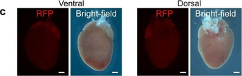
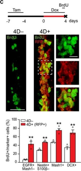
Transgenic model and effects of acute 4D overexpression on NSC and progenitors of the SVZDrawings of the nestinCreERT2, ROSA26rtTA‐flox, and tet4D‐RFP alleles of the 4D line. From top to bottom: experimental design of 4D induction, fluorescence pictures of the SVZ of a 4D+ mouse and quantification of the proportion of RFP− (black) and RFP+ (red) progenitors among B1, C, and A cells identified with markers as indicated.From top to bottom: experimental design, fluorescence pictures of the SVZ of a 4D− (left) and 4D+ (right and insets magnified) mice, and quantification of the proportion of BrdU + among B1, C, and A cells (identified as in B). Note that in 4D+ mice quantification was restricted to the RFP+ subpopulation (red bars).Quantification of the absolute number of B1, C, and A cells (identified as in B) in the SVZ of 4D− (white) and 4D+ (black) mice regardless of RFP expression (RFP+/−). Data information: (B–D) Mean ± SEM; *P 423 cells for each quantification. Scale bar = 50 µm (B and C) or 20 µm (inset in C).

Transgenic model and effects of acute 4D overexpression on NSC and progenitors of the SVZDrawings of the nestinCreERT2, ROSA26rtTA‐flox, and tet4D‐RFP alleles of the 4D line. From top to bottom: experimental design of 4D induction, fluorescence pictures of the SVZ of a 4D+ mouse and quantification of the proportion of RFP− (black) and RFP+ (red) progenitors among B1, C, and A cells identified with markers as indicated.From top to bottom: experimental design, fluorescence pictures of the SVZ of a 4D− (left) and 4D+ (right and insets magnified) mice, and quantification of the proportion of BrdU + among B1, C, and A cells (identified as in B). Note that in 4D+ mice quantification was restricted to the RFP+ subpopulation (red bars).Quantification of the absolute number of B1, C, and A cells (identified as in B) in the SVZ of 4D− (white) and 4D+ (black) mice regardless of RFP expression (RFP+/−). Data information: (B–D) Mean ± SEM; *P 423 cells for each quantification. Scale bar = 50 µm (B and C) or 20 µm (inset in C).
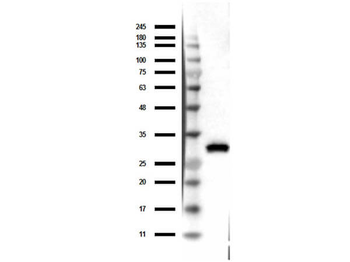
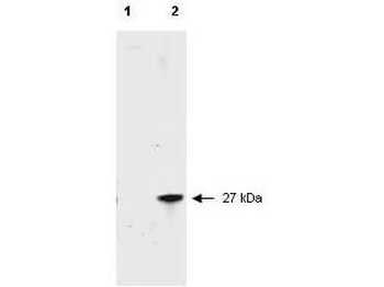
Western Blot of Mouse Anti-RFP Antibody. Lane 1: Opal Prestain Molecular weight. Lane 2: 50 ng of RFP. Primary Antibody: Mouse Anti-RFP at 1 µg/ml overnight at 2-8°C. Secondary Antibody: Rabbit Anti-Mouse HRP (p/n orb347506) at 1:40000 for 30 mins at RT. Block: BlockOut Universal blocking buffer (p/n orb348644). Expect ~27kDa.
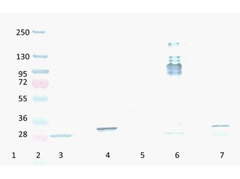
Western Blot of Mouse Anti-RFP antibody. Lane 1: YFP protein. Lane 2: Prestained Molecular Weight Marker. Lane 3: Reduced RFP control Protein. Lane 4: Reduced mCherry. Lane 5: GFP protein. Lane 6: Non-Reduced RFP control Protein. Lane 7: Non-Reduced mCherry. Load: 300 ng per lane. Primary antibody: RFP antibody at 1:2000 in orb348637 for 3 hours at RT. Secondary antibody: HRP anti-Mouse secondary antibody at 1:10000 in orb348637 for 60 min at RT. Substrate: orb348656 for 20 min. Predicted/Observed size: ~27 kDa.
Quick Database Links
Gene Symbol
DsRed
UniProt
UniProt Details
− No UniProt data available
Documents Download
Datasheet
Product Information
Request a Document
Protocol Information
WB
Western Blot (IB, immunoblot)
ELISA
Enzyme-linked Immunosorbent Assay (EIA)
DsRed Antibody (orb344411)

- 0.0
Based on 0 reviews
Participating in our Biorbyt product reviews program enables you to support fellow scientists by sharing your firsthand experience with our products.
Login to Submit a ReviewAvailable Sizes
Select a size below
Free Secondary Antibody (20 ul)0/0
Please add an antibody product to your cart first.