You have no items in your shopping cart.
Description
Research Area
Cell Biology
Images & Validation
−Item 1 of 2
| Tested Applications | ELISA, IHC, WB |
|---|---|
| Dilution Range | ELISA: 1:4,000 - 1:20,000, IHC: 1:500 - 1:3,000, WB: 1:500 - 1:3,000 |
| Reactivity | Human |
| Application Notes |
Key Properties
−| Antibody Type | Primary Antibody |
|---|---|
| Host | Rabbit |
| Clonality | Polyclonal |
| Isotype | IgG |
| Immunogen | This affinity purified antibody was prepared from whole rabbit serum produced by repeated immunizations with a recombinant protein corresponding to amino acids 1-363 of human RFFL protein. |
| Target | RFFL |
| Purity | This protein A purified antibody is directed against human RFFL protein. The product was purified from monospecific antiserum by protein A chromatography followed by exhaustive dialysis against the buffer stated above. |
| Conjugation | Unconjugated |
Storage & Handling
−| Storage | Store vial at 4° C prior to restoration. For extended storage aliquot contents and freeze at -20° C or below. Avoid cycles of freezing and thawing. Centrifuge product if not completely clear after standing at room temperature. This product is stable for several weeks at 4° C as an undiluted liquid. Dilute only prior to immediate use. |
|---|---|
| Form/Appearance | Lyophilized |
| Buffer/Preservatives | Preservative: 0.01% (w/v) Sodium Azide. Stabilizer: None; Buffer: 0.02 M Potassium Phosphate, 0.15 M Sodium Chloride, pH 7.2 |
| Concentration | 5.0 mg/mL |
| Expiration Date | 12 months from date of receipt. |
| Disclaimer | For research use only |
Alternative Names
−rabbit anti-RFFL antibody, Rififylin, RING finger and FYVE-like domain-containing protein 1, FYVE-RING finger protein, Sakura, Fring, Caspases-8 and -10-associated RING finger protein 2, CARP-2, Caspase regulator CARP2, RING finger protein 189 and RING finger protein 34-like
Similar Products
−- Item 1 of 1
RING finger protein 189 Rabbit Polyclonal Antibody [orb100410]
WB
Bovine, Canine, Porcine, Rabbit, Rat, Sheep
Human, Mouse
Rabbit
Polyclonal
Unconjugated
50 μl, 100 μl, 200 μl - Item 1 of 2
- Item 1 of 1
- Item 1 of 1
- Item 1 of 1
RFFL Rabbit Polyclonal Antibody [orb630641]
ELISA, WB
Human, Mouse, Rat
Rabbit
Polyclonal
Unconjugated
50 μg, 100 μg


Quality Guarantee
Explore bioreagents carefree to elevate your research. All our products are rigorously tested for performance. If a product does not perform as described on its datasheet, our scientific support team will provide expert troubleshooting, a prompt replacement, or a refund. For full details, please see our Terms & Conditions and Buying Guide. Contact us at [email protected].
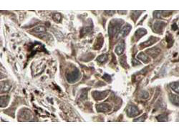
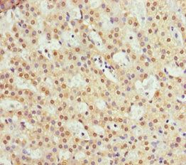
Biorbyt's Affinity Purified anti-RFFL antibody shows strong cytoplasmic and membranous staining of tumor cells in cancerous human liver tissue. Tissue was formalin-fixed and paraffin embedded. Brown color indicates presence of protein, blue color shows cell nuclei.
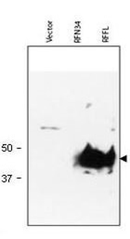
Western blot using Biorbyt's Protein A Purified anti-RFFL antibody shows detection of RFFL (arrowhead) in lysate. Lanes correspond to empty vector 293T cell lysate (mock, left); RNF34 transfected lysate (middle) and RFFL transfected lysate (right), are shown using 20 µl of lysate per lane. Lysates were prepared from equivalent numbers of cells. Data presented demonstrate that this reagent is specific for RFFL. After SDS-PAGE and transfer, the membrane was probed with the primary antibody diluted to 1:1000 using 5% BLOTTO, 0.1% Tween-20 in PBS as the diluent. Incuba-tion occurred for 1 h at room temperature.
Documents Download
Datasheet
Product Information
Request a Document
Protocol Information
WB
Western Blot (IB, immunoblot)
IHC
Immunohistochemistry
ELISA
Enzyme-linked Immunosorbent Assay (EIA)
RFFL Antibody (orb344634)

- 0.0
Based on 0 reviews
Participating in our Biorbyt product reviews program enables you to support fellow scientists by sharing your firsthand experience with our products.
Login to Submit a ReviewAvailable Sizes
Select a size below
Free Secondary Antibody (20 ul)0/0
Please add an antibody product to your cart first.