You have no items in your shopping cart.
Description
Research Area
Cancer Biology, Cardiovascular Research, Immunology & Inflammation, Neuroscience
Images & Validation
−Item 1 of 3
| Tested Applications | ChIP, IHC, IP, WB |
|---|---|
| Dilution Range | WB - 1:2,000 - 1:10,000; IP - 2 - 5 µg/mg lysate; IHC - 1:500 - 1:2,000. Epitope retrieval with citrate buffer pH 6.0 is recommended for FFPE tissue sections. |
| Reactivity | Human, Mouse |
| Application Notes |
Key Properties
−| Antibody Type | Primary Antibody |
|---|---|
| Host | Rabbit |
| Clonality | Polyclonal |
| Isotype | IgG |
| Immunogen | Between 501 and C-terminus |
| Target | RelA |
| Purification | Antigen Affinity Purified |
| Conjugation | Unconjugated |
Storage & Handling
−| Storage | 2 - 8°C |
|---|---|
| Form/Appearance | Liquid |
| Buffer/Preservatives | Tris-citrate/phosphate buffer, pH 7 to 8 containing 0.09% Sodium Azide |
| Concentration | 1000 µg/ml |
| Expiration Date | 12 months from date of receipt. |
| Disclaimer | For research use only |
Alternative Names
−NF-kappa-B p65delta3; NF-kappa-B transcription factor p65; NFKB3; nuclear factor NF-kappa-B p65 subunit; nuclear factor of kappa light polypeptide gene enhancer in B-cells 3; p65; transcription factor p65; v-rel avian reticuloendotheliosis viral oncogene homolog A; v-rel reticuloendotheliosis viral oncogene homolog A
Similar Products
−- Item 1 of 15
NFKB p65 Rabbit Polyclonal Antibody [orb11118]
FC, ICC
Bovine, Canine, Equine, Gallus, Porcine, Rabbit, Rat, Sheep, Zebrafish
Human, Mouse
Rabbit
Polyclonal
Unconjugated
50 μl, 100 μl, 200 μl - Item 1 of 9
NFKB p65 Rabbit Polyclonal Antibody [orb312399]
FC, ICC, IF, IHC-Fr, IHC-P, KO/KD Validated, WB
Bovine, Canine, Porcine
Human, Mouse, Rat
Rabbit
Polyclonal
Unconjugated
50 μl, 100 μl, 200 μl - Item 1 of 8
Phospho-NFKB p65 (Ser468) Rabbit Polyclonal Antibody [orb6503]
FC, ICC, IF, IHC-Fr, IHC-P, WB
Bovine, Canine, Equine, Porcine
Human, Mouse, Rat
Rabbit
Polyclonal
Unconjugated
50 μl, 100 μl, 200 μl - Item 1 of 6
Phospho-NFKB p65 (Ser536) Rabbit Polyclonal Antibody [orb6504]
WB
Human, Monkey, Rat
Mouse
Rabbit
Polyclonal
Unconjugated
50 μl, 100 μl, 200 μl - Item 1 of 8
NFKB p65 Recombinant Rabbit Monoclonal Antibody [orb608066]
FC, ICC, IF, IHC-Fr, IHC-P, KO/KD Validated, WB
Zebrafish
Human, Mouse, Rat
Rabbit
Recombinant
Unconjugated
50 μl, 100 μl, 25 μl


Quality Guarantee
Explore bioreagents carefree to elevate your research. All our products are rigorously tested for performance. If a product does not perform as described on its datasheet, our scientific support team will provide expert troubleshooting, a prompt replacement, or a refund. For full details, please see our Terms & Conditions and Buying Guide. Contact us at [email protected].
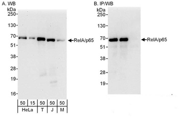
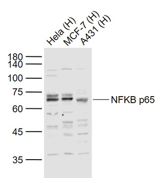
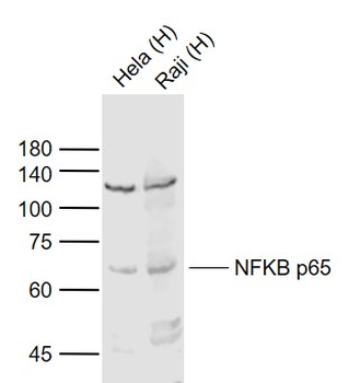
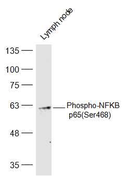
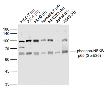
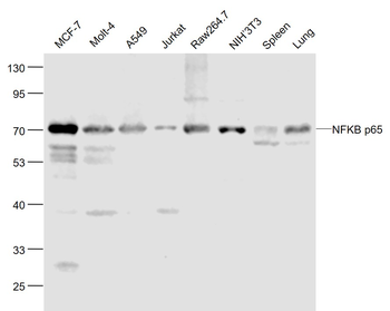
Detection of human and mouse RelA/p65 by western blot (h&m) and immunoprecipitation (h). Samples: Whole cell lysate from HeLa (15 and 50 µg for WB; 1 mg for IP, 20% of IP loaded) , HEK293T (T; 50 µg) , Jurkat (J; 50 µg) and mouse NIH 3T3 (M; 50 µg) cells. Antibodies: Affinity purified rabbit anti-RelA/p65 antibody orb1527723 used for WB at 0.1 µg/ml (A) and 0.4 µg/ml (B) and used for IP at 3 µg/mg lysate. RelA/p65 was also immunoprecipitated by a previous lot of this antibody. Detection: Chemiluminescence with exposure times of 30 seconds (A) and 10 seconds (B).
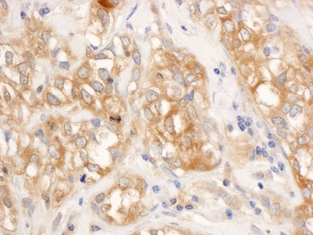
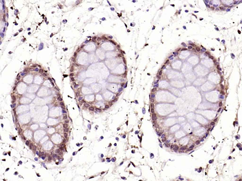
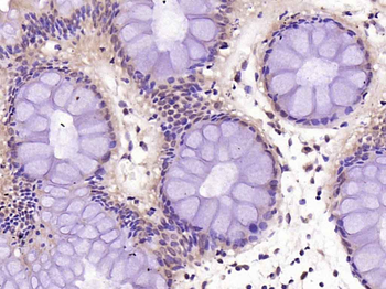
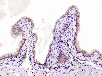
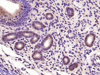
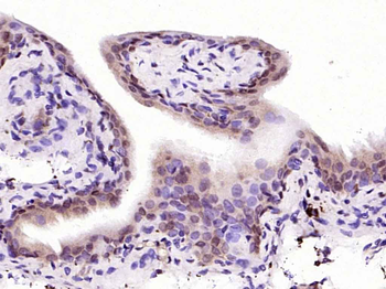
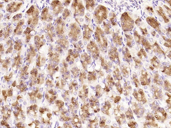
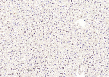
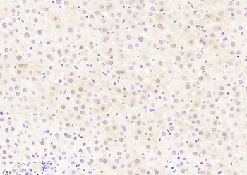
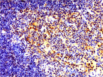
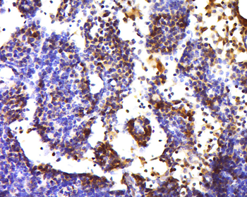
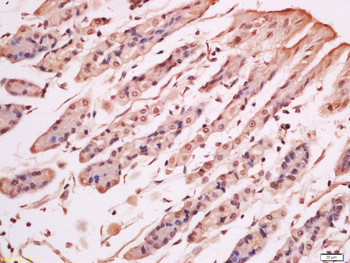
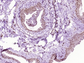
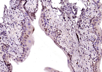
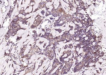
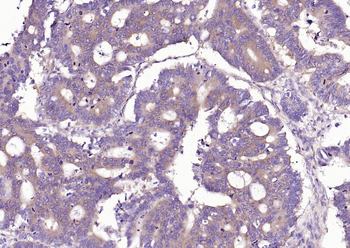
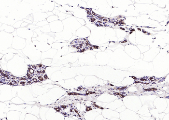
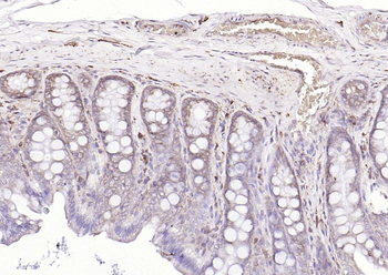
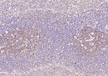
Detection of human RelA/p65 by immunohistochemistry.Sample: FFPE section of human breast carcinoma. Antibody: Affinity purified rabbit anti-RelA/p65 (Cat. No. orb1527723) used at a dilution of 1: 1, 000 (1µg/ml). Detection: DAB

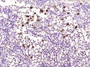
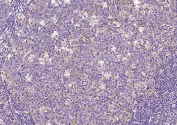
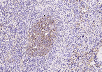
Localization of RelA Binding Sites by ChIP-sequencing. Chromatin from human ependymoma tumor was immunoprecipitated with anti-RelA antibody orb1527723 and analyzed by DNA sequencing. The figure illustrates the peak distribution of RelA binding within a 250 Kb region of chromosome 19 as detected using anti-RelA orb1527723.
Quick Database Links
UniProt Details
− No UniProt data available
NCBI Reference Sequences
−Associated Accession Numbers
Curated reference sequences for the gene transcript and protein product| Protein | NP_068810.3 |
|---|
Documents Download
Datasheet
Product Information
Request a Document
Protocol Information
WB
Western Blot (IB, immunoblot)
IHC
Immunohistochemistry
IP
Immunoprecipitation
ChIP
Chromatin Immunoprecipitation
Rabbit RelA Antibody (orb1527723)

- 0.0
Based on 0 reviews
Participating in our Biorbyt product reviews program enables you to support fellow scientists by sharing your firsthand experience with our products.
Login to Submit a ReviewAvailable Sizes
Select a size below
Choose Conjugation or Carrier Free Version
Free Secondary Antibody (20 ul)0/0
Please add an antibody product to your cart first.