You have no items in your shopping cart.
Description
Research Area
Neuroscience
Images & Validation
−Item 1 of 2
| Tested Applications | IHC, IP, WB |
|---|---|
| Dilution Range | WB - 1:2,000 - 1:10,000; IP - 10 µg/mg lysate; IHC - 1:500 - 1:2,000. Epitope retrieval with citrate buffer pH 6.0 is recommended for FFPE tissue sections. |
| Reactivity | Human |
| Application Notes |
Key Properties
−| Antibody Type | Primary Antibody |
|---|---|
| Host | Rabbit |
| Clonality | Polyclonal |
| Isotype | IgG |
| Immunogen | Between 25 and 75 |
| Target | RAI1 |
| Purification | Antigen Affinity Purified |
| Conjugation | Unconjugated |
Storage & Handling
−| Storage | 2 - 8°C |
|---|---|
| Form/Appearance | Liquid |
| Buffer/Preservatives | Tris-citrate/phosphate buffer, pH 7 to 8 containing 0.09% Sodium Azide |
| Concentration | 1000 µg/ml |
| Expiration Date | 12 months from date of receipt. |
| Disclaimer | For research use only |
Alternative Names
−retinoic acid-induced protein 1; SMCR; Smith-Magenis syndrome chromosome region; SMS
Similar Products
−- Item 1 of 1
RAI1 Rabbit Polyclonal Antibody [orb630495]
ELISA, WB
Human, Mouse, Rat
Rabbit
Polyclonal
Unconjugated
50 μg, 100 μg - Item 1 of 1
RAI1 Rabbit Polyclonal Antibody [orb412855]
WB
Human, Mouse, Rat
Rabbit
Polyclonal
Unconjugated
50 μl, 100 μl, 200 μl, 30 μl - Item 1 of 1
DXO Rabbit Polyclonal Antibody [orb581564]
WB
Bovine, Canine, Equine, Guinea pig, Mouse, Porcine, Rabbit, Rat
Human
Rabbit
Polyclonal
Unconjugated
100 μl 
RAI1 Rabbit Polyclonal Antibody (HRP) [orb470272]
ELISA, IHC-Fr, IHC-P, WB
Human, Mouse, Rat
Rabbit
Polyclonal
HRP
100 μlRAI1 Rabbit Polyclonal Antibody (Cy5) [orb917421]
ICC, IF
Human, Mouse, Rat
Rabbit
Polyclonal
Cy5
100 μl
Quality Guarantee
Explore bioreagents carefree to elevate your research. All our products are rigorously tested for performance. If a product does not perform as described on its datasheet, our scientific support team will provide expert troubleshooting, a prompt replacement, or a refund. For full details, please see our Terms & Conditions and Buying Guide. Contact us at [email protected].

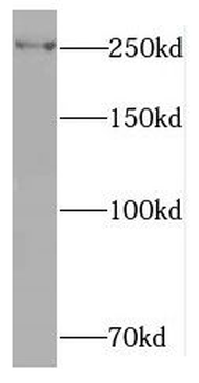
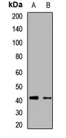
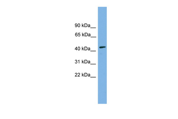
Detection of human RAI1 by western blot and immunoprecipitation. Samples: Whole cell lysate (5, 15 and 50 µg for WB; 1 mg for IP, 20% of IP loaded) from HeLa cells. Antibodies: Affinity purified rabbit anti-RAI1 antibody orb1526840 used for WB at 0.1 µg/ml (A) and 1 µg/ml (B) and used for IP at 10 µg/mg lysate. RAI1 was also immunoprecipitated by rabbit anti-RAI1 antibody, which recognizes a downstream epitope. Detection: Chemiluminescence with exposure times of 3 minutes (A) and 30 seconds (B).
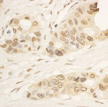
Detection of human RAI1 by immunohistochemistry.Sample: FFPE section of human ovarian carcinoma. Antibody: Affinity purified rabbit anti-RAI1 (Cat. No. orb1526840) used at a dilution of 1: 1, 000 (1µg/ml). Detection: DAB
Quick Database Links
UniProt Details
− No UniProt data available
NCBI Reference Sequences
−Associated Accession Numbers
Curated reference sequences for the gene transcript and protein product| Protein | NP_109590.3 |
|---|
Documents Download
Datasheet
Product Information
Request a Document
Protocol Information
WB
Western Blot (IB, immunoblot)
IHC
Immunohistochemistry
IP
Immunoprecipitation
Rabbit RAI1 Antibody (orb1526840)

- 0.0
Based on 0 reviews
Participating in our Biorbyt product reviews program enables you to support fellow scientists by sharing your firsthand experience with our products.
Login to Submit a ReviewAvailable Sizes
Select a size below
Choose Conjugation or Carrier Free Version
Free Secondary Antibody (20 ul)0/0
Please add an antibody product to your cart first.