You have no items in your shopping cart.
Description
Research Area
Signal Transduction
Images & Validation
−
Item 1 of 4
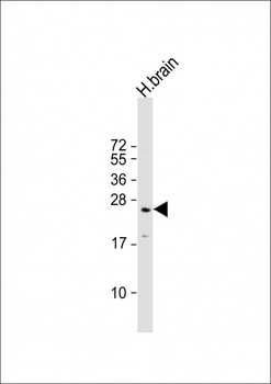
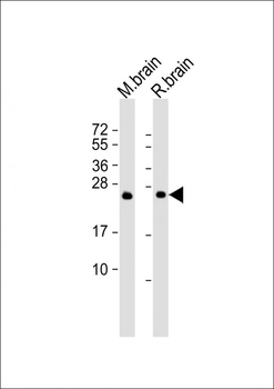
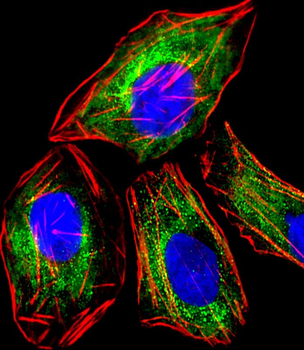
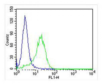
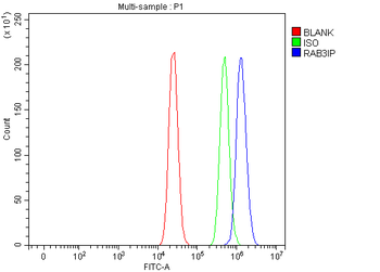
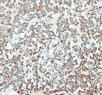
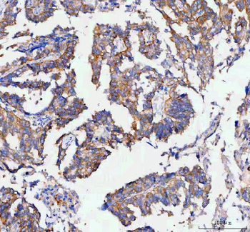
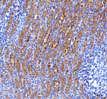
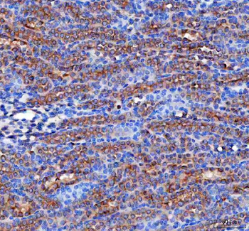
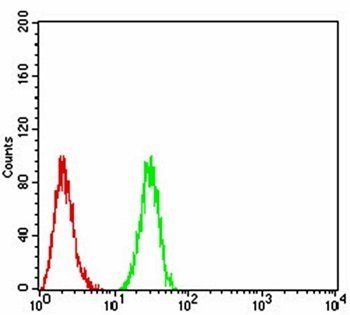
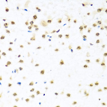
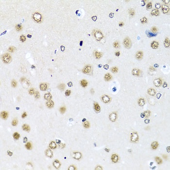
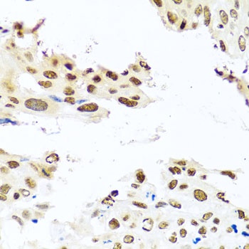
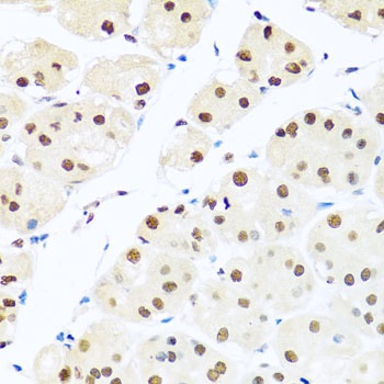
| Tested Applications | FC, IF, WB |
|---|---|
| Dilution Range | IF - 1:25, WB - 1:2000, FC - 1:25 |
| Reactivity | Human, Mouse, Rat |
Key Properties
−| Host | Mouse |
|---|---|
| Clonality | Monoclonal |
| Isotype | IgG1,k |
| Clone No. | B3753EV784X36X79 |
| Immunogen | This RAB3A antibody is generated from a mouse immunized with a recombinant protein. |
| Target | RAB3A (HGNC:9777) |
| Molecular Weight | 24984 Da |
| Conjugation | Unconjugated |
Storage & Handling
−| Storage | Maintain refrigerated at 2-8°C for up to 2 weeks. For long term storage store at -20°C in small aliquots to prevent freeze-thaw cycles |
|---|---|
| Form/Appearance | Purified monoclonal antibody supplied in PBS with 0.09% (W/V) sodium azide. This antibody is purified through a protein G column, followed by dialysis against PBS. |
| Expiration Date | 12 months from date of receipt. |
| Disclaimer | For research use only |
Alternative Names
−Ras-related protein Rab-3A, RAB3A
Similar Products
−- Item 1 of 6
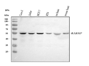
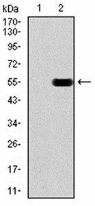
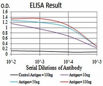
RAB3IP Rabbit Polyclonal Antibody [orb1819445]
ELISA, FC, IHC, WB
Human, Mouse, Rat
Rabbit
Polyclonal
Unconjugated
100 μg - Item 1 of 1
Rat Ras-Related Protein Rab-3A (RAB3A) ELISA Kit [orb1736751]
Rat
78.13-5000 pg/mL
32 pg/mL
48 T, 96 T - Item 1 of 4
- Item 1 of 5


Quality Guarantee
Explore bioreagents carefree to elevate your research. All our products are rigorously tested for performance. If a product does not perform as described on its datasheet, our scientific support team will provide expert troubleshooting, a prompt replacement, or a refund. For full details, please see our Terms & Conditions and Buying Guide. Contact us at [email protected].
Quick Database Links
Gene Symbol
RAB3A (HGNC:9777)
UniProt
UniProt Details
− No UniProt data available
Protocol Information
WB
Western Blot (IB, immunoblot)
FC
Flow Cytometry
IF
Immunofluorescence
Available Sizes
Select a size below
Choose Conjugation or Carrier Free Version
Free Secondary Antibody (20 ul)0/0
Please add an antibody product to your cart first.