You have no items in your shopping cart.
Featured
Description
Research Area
Metabolism Research
Images & Validation
−Item 1 of 3
| Tested Applications | AM, ICC, IF, IHC, WB |
|---|---|
| Dilution Range | WB (1:250), IHC (1:1000), ICC/IF (1:100) |
| Reactivity | Bovine, Human, Mouse, Rat |
| Application Notes |
Key Properties
−| Host | Mouse |
|---|---|
| Clonality | Monoclonal |
| Isotype | IgG2a |
| Clone No. | 6G6 |
| Immunogen | Recombinant rat PSD-95 |
| Target | PSD95 |
| Molecular Weight | 75kDa |
| Purification | Protein A Purified |
| Conjugation | RPE |
Storage & Handling
−| Storage | Conjugated antibodies should be stored according to the product label |
|---|---|
| Buffer/Preservatives | 95.46mM Phosphate, 2.48mM MES and 2mM EDTA |
| Concentration | 1 mg/ml |
| Expiration Date | 12 months from date of receipt. |
| Disclaimer | For research use only |
Alternative Names
−PSD 95, PSD-95, PSD95, DLG4, SAP90, Synapse-associated protein 90, Postsynaptic density protein 95, Disks large homolog 4
Similar Products
−- Item 1 of 4
PSD95 Antibody (RPE) [orb147029]
AM, ICC, IF, IHC, WB
Bovine, Human, Mouse, Rat
Mouse
Monoclonal
RPE
100 μg 
Quality Guarantee
Explore bioreagents carefree to elevate your research. All our products are rigorously tested for performance. If a product does not perform as described on its datasheet, our scientific support team will provide expert troubleshooting, a prompt replacement, or a refund. For full details, please see our Terms & Conditions and Buying Guide. Contact us at [email protected].
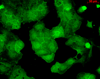
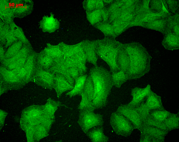
Immunocytochemistry/Immunofluorescence analysis using Mouse Anti-PSD95 Monoclonal Antibody, Clone 6G6. Tissue: HaCaT cells. Species: Human. Fixation: Cold 100% methanol for 10 minutes at -20°C. Primary Antibody: Mouse Anti-PSD95 Monoclonal Antibody at 1:100 for 1 hour at RT. Secondary Antibody: FITC Goat Anti-Mouse (green) at 1:50 for 1 hour at RT. Localization: Junction staining.
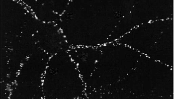
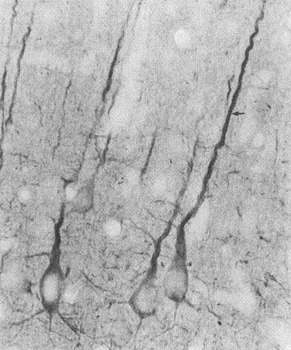
Immunocytochemistry/Immunofluorescence analysis using Mouse Anti-PSD95 Monoclonal Antibody, Clone 6G6. Tissue: dissociated hippocampal neurons. Species: Rat. Fixation: Cold 4% paraformaldehyde/0.2% glutaraldehyde in 0.1M sodium phosphate buffer. Primary Antibody: Mouse Anti-PSD95 Monoclonal Antibody at 1:1000 for 12 hours at 4°C. Secondary Antibody: FITC Goat Anti-Mouse IgG (green) at 1:50 for 30 minutes at RT. Magnification: 10X.
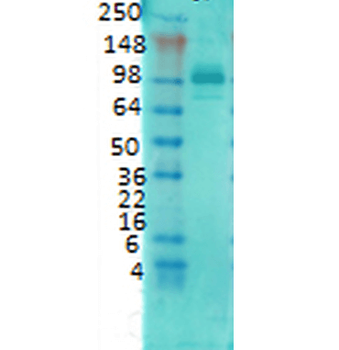
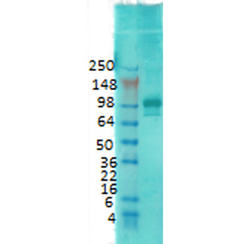
Western Blot analysis of Rat brain membrane lysate showing detection of PSD95 protein using Mouse Anti-PSD95 Monoclonal Antibody, Clone 6G6. Primary Antibody: Mouse Anti-PSD95 Monoclonal Antibody at 1:1000.
Quick Database Links
UniProt Details
− No UniProt data available
NCBI Gene Details
− No NCBI Gene data available
NCBI Reference Sequences
−Associated Accession Numbers
Curated reference sequences for the gene transcript and protein product| Protein | NP_062567.1 |
|---|
Documents Download
Datasheet
Product Information
Request a Document
Protocol Information
WB
Western Blot (IB, immunoblot)
IHC
Immunohistochemistry
IF
Immunofluorescence
ICC
Immunocytochemistry
PSD95 Antibody (RPE) (orb147012)

- 0.0
Based on 0 reviews
Participating in our Biorbyt product reviews program enables you to support fellow scientists by sharing your firsthand experience with our products.
Login to Submit a ReviewAvailable Sizes
Select a size below
Choose Conjugation or Carrier Free Version
Free Secondary Antibody (20 ul)0/0
Please add an antibody product to your cart first.