You have no items in your shopping cart.
On Promotion
Category
Tested Applications
Predicted Reactivity
Conjugation
Biologically Active
Featured Product
Search results for: 'GM-CSF'
- Active
 Item 1 of 2Human GM-CSF Protein [orb257510]Active
Unconjugated
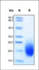
95%
14.5 kDa
Ala 18 - Glu 144
Native
50 μg, 1 mg, 20 μg - Featured
 ActiveItem 1 of 2FeaturedActive
Greater than 95% as determined by SDS-PAGE.
50.9 kDa
Mammalian cell
Homo sapiens (Human)
17-443aa
Partial
C-terminal 6xHis-tagged
100 μg, 20 μg, 1 mg - Item 1 of 1
46.9 kDa
Human
C-Myc/DDK
20 μg - FeaturedItem 1 of 2Featured
Unconjugated
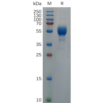
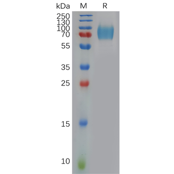
The purity of the protein is greater than 95% as determined by SDS-PAGE and Coomassie blue staining.
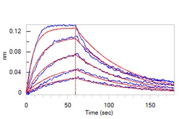
The protein has a predicted molecular mass of 60.6 kDa after removal of the signal peptide.The apparent molecular mass of GM-CSFR-hFc is approximately 70-130 kDa due to glycosylation.
Mammalian
GM-CSFR(Glu23-Gly320) hFc(Glu99-Ala330)
C-Human Fc Tag
10 μg, 100 μg, 50 μg - FeaturedItem 1 of 2Featured
Unconjugated
The purity of the protein is greater than 95% as determined by SDS-PAGE and Coomassie blue staining.
The protein has a predicted molecular mass of 40.6 kDa after removal of the signal peptide.The apparent molecular mass of GM-CSF-hFc is approximately 35-70 kDa due to glycosylation.
Mammalian
GM-CSF(Ala18-Glu144) hFc(Glu99-Ala330)
C-Human Fc Tag
100 μg, 50 μg, 10 μg - Item 1 of 1
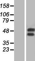
46.2 kDa
Human
C-Myc/DDK
20 μg - Item 1 of 1
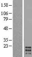
16.3 kDa
Human
C-Myc/DDK
20 μg - Item 1 of 1
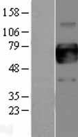
46.9 kDa
Human
C-Myc/DDK
100 μg - Item 1 of 1
36.3 kDa
Human
C-Myc/DDK
20 μg - ActiveItem 1 of 1Active
Unconjugated
95%
60.9 kDa
Glu 23 - Gly 320
C-Fc
100 μg, 1 mg