You have no items in your shopping cart.
Featured
Description
Research Area
Epigenetics & Chromatin, Signal Transduction
Images & Validation
−Item 1 of 3
| Tested Applications | ICC, IF, WB |
|---|---|
| Dilution Range | WB (1:1000), ICC/IF (1:100) |
| Reactivity | Human |
| Application Notes |
Key Properties
−| Host | Mouse |
|---|---|
| Clonality | Monoclonal |
| Isotype | IgG1 |
| Clone No. | 12F7 |
| Immunogen | Full length human PP5 protein |
| Target | PP5 |
| Molecular Weight | 58kDa |
| Purification | Protein G Purified |
| Conjugation | APC |
Storage & Handling
−| Storage | Conjugated antibodies should be stored according to the product label |
|---|---|
| Buffer/Preservatives | 95.46mM Phosphate, 2.48mM MES and 2mM EDTA |
| Concentration | 1 mg/ml |
| Expiration Date | 12 months from date of receipt. |
| Disclaimer | For research use only |
Alternative Names
−PPP5, PPT, Protein phosphatase T, Serine/threonine protein phosphatase 5, PPP5C
Similar Products
−- Item 1 of 5
FKBP52 Antibody (APC) [orb147210]
ICC, IF, IHC, IP, WB
Canine, Hamster, Human, Mouse, Rat
Mouse
Monoclonal
APC
100 μg - Item 1 of 2
FKBP51 Antibody (APC) [orb147193]
ICC, IF, WB
Canine, Hamster, Human, Mouse, Rabbit, Rat
Mouse
Monoclonal
APC
100 μg 
TFPI-2 Rabbit Polyclonal Antibody (APC) [orb1005805]
IF
Bovine, Mouse
Human, Rat
Rabbit
Polyclonal
APC
100 μl
Quality Guarantee
Explore bioreagents carefree to elevate your research. All our products are rigorously tested for performance. If a product does not perform as described on its datasheet, our scientific support team will provide expert troubleshooting, a prompt replacement, or a refund. For full details, please see our Terms & Conditions and Buying Guide. Contact us at [email protected].
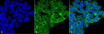
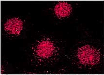
Immunocytochemistry/Immunofluorescence analysis using Mouse Anti-PP5 Monoclonal Antibody, Clone 12F7. Tissue: Embryonic kidney epithelial cell line (HEK293). Species: Human. Fixation: 2% Formaldehyde for 20 min at RT. Primary Antibody: Mouse Anti-PP5 Monoclonal Antibody at 1:50 for 1 hour at RT. Secondary Antibody: Alexa Fluor 488 Goat Anti-Mouse (green) at 1:100 for 1 hour at RT. Counterstain: DAPI (blue) nuclear stain. Magnification: 63x.
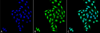
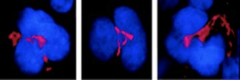
Immunocytochemistry/Immunofluorescence analysis using Mouse Anti-PP5 Monoclonal Antibody, Clone 12F7. Tissue: Cervical cancer cell line (HeLa). Species: Human. Fixation: 4% Formaldehyde for 15 min at RT. Primary Antibody: Mouse Anti-PP5 Monoclonal Antibody at 1:100 for 60 min at RT. Secondary Antibody: Goat Anti-Mouse ATTO 488 at 1:100 for 60 min at RT. Counterstain: DAPI (blue) nuclear stain at 1:5000 for 5 min RT. Localization: Nucleus, Cytoplasm. Magnification: 40X.
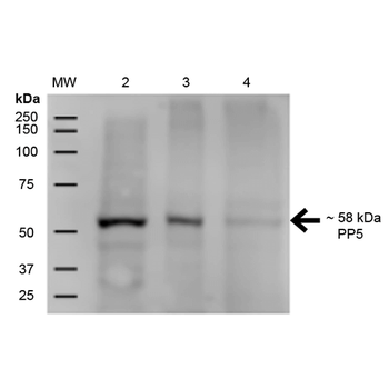
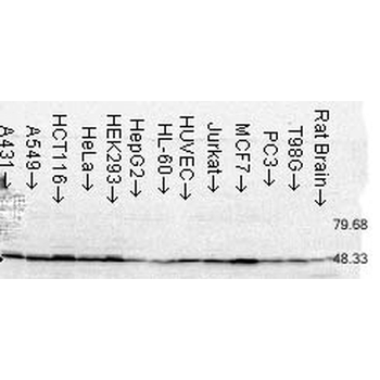
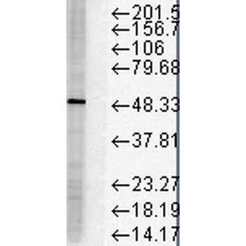
Western Blot analysis of Human A431, HEK293, and Jurkat cell lysates showing detection of ~58 kDa PP5 protein using Mouse Anti-PP5 Monoclonal Antibody, Clone 12F7. Lane 1: MW Ladder. Lane 2: Human A431 (15 μg). Lane 3: Human HEK293 (15 μg). Lane 4: Human Jurkat (15 μg). Load: 15 μg. Block: 5% Skim Milk for 1 hour at RT. Primary Antibody: Mouse Anti-PP5 Monoclonal Antibody at 1:500 for 1 hour at RT. Secondary Antibody: Goat Anti-Mouse IgG: HRP at 1:200 for 1 hour at RT. Color Development: ECL solution for 6 min at RT. Predicted/Observed Size: ~58 kDa.
Quick Database Links
UniProt Details
− No UniProt data available
NCBI Gene Details
− No NCBI Gene data available
NCBI Reference Sequences
−Associated Accession Numbers
Curated reference sequences for the gene transcript and protein product| Protein | NP_006238.1 |
|---|
Documents Download
Datasheet
Product Information
Request a Document
Protocol Information
WB
Western Blot (IB, immunoblot)
IF
Immunofluorescence
ICC
Immunocytochemistry
PP5 Antibody (APC) (orb377030)

- 0.0
Based on 0 reviews
Participating in our Biorbyt product reviews program enables you to support fellow scientists by sharing your firsthand experience with our products.
Login to Submit a ReviewAvailable Sizes
Select a size below
Choose Conjugation or Carrier Free Version
Free Secondary Antibody (20 ul)0/0
Please add an antibody product to your cart first.