You have no items in your shopping cart.
Featured
Description
Research Area
Cancer Biology
Images & Validation
−Item 1 of 7
| Tested Applications | IF, IHC-Fr, IHC-P |
|---|---|
| Dilution Range | IHC-P=1:100-500, IHC-F=1:100-500, IF=1:100-500 |
| Reactivity | Human, Mouse, Rat |
| Predicted Reactivity | Bovine, Canine, Equine, Porcine, Rabbit, Sheep |
Related Conjugates & Formulations
−Key Properties
−| Antibody Type | Primary Antibody |
|---|---|
| Host | Rabbit |
| Clonality | Polyclonal |
| Isotype | IgG |
| Immunogen | KLH conjugated synthetic peptide derived from human PI3 kinase p85 subunit alpha (501-600/724aa) |
| Target | PIK3R1 |
| Molecular Weight | 80 kDa |
| Purification | Affinity purified by Protein A |
| Conjugation | Unconjugated |
Storage & Handling
−| Storage | Maintain refrigerated at 2-8°C for up to 2 weeks. For long term storage store at -20°C in small aliquots to prevent freeze-thaw cycles. |
|---|---|
| Form/Appearance | Liquid |
| Buffer/Preservatives | 0.01M TBS (pH7.4) with 1% rAlbumin, 0.02% Proclin300 and 50% Glycerol. |
| Concentration | 1mg/ml |
| Expiration Date | 12 months from date of receipt. |
| Disclaimer | For research use only |
Alternative Names
−AGM7; GRB1; IMD36; p85; p85-ALPHA; p85alpha; PI3K; p50alpha; p55alpha; PI3KA; P85A_HUMAN; PIK3R1; PI3-kinase regulatory subunit alpha; PI3K regulatory subunit alpha; PtdIns-3-kinase regulatory subunit alpha; Phosphatidylinositol 3-kinase 85 kDa regulatory subunit alpha (PI3-kinase subunit p85-alpha | PtdIns-3-kinase regulatory subunit p85-alpha); P85A_MOUSE; P85A_RAT; phosphoinositide-3-kinase regulatory subunit 1; phosphoinositide-3-kinase, regulatory subunit 1 (alpha); phosphoinositide-3-kinase regulatory subunit alpha; growth factor receptor bound 1; PI3 kinase-associated p85
Similar Products
−- Item 1 of 2
Phospho-PI3 kinase p85 alpha + gamma (Tyr467 + Tyr199) Rabbit Polyclonal Antibody [orb106105]
FC, IF, IHC-Fr, IHC-P
Bovine, Canine, Equine, Mouse, Porcine, Rabbit
Human, Rat
Rabbit
Polyclonal
Unconjugated
50 μl, 100 μl, 200 μl - Item 1 of 8
PI 3 Kinase p85 alpha/PIK3R1 Rabbit Polyclonal Antibody [orb763138]
ELISA, FC, ICC, IF, IHC, WB
Human, Mouse, Rat
Rabbit
Polyclonal
Unconjugated
100 μg - Item 1 of 5
PI3KR1 Antibody (N-term L11) [orb38864]
FC, IF, IHC-P, WB
Mouse
Human, Rat
Rabbit
Polyclonal
Unconjugated
50 μl, 100 μl - Item 1 of 7
PI 3-kinase p85/p55 (phospho Tyr467/199) Polyclonal Antibody [orb1415313]
IF, IHC-P, WB
Human, Mouse, Rat
Rabbit
Polyclonal
Unconjugated
100 μl - Item 1 of 4
PI 3-kinase p85α rabbit pAb Antibody [orb766074]
ELISA, IF, IHC, WB
Human, Mouse, Rat
Polyclonal
Unconjugated
50 μl, 100 μl

Quality Guarantee
Explore bioreagents carefree to elevate your research. All our products are rigorously tested for performance. If a product does not perform as described on its datasheet, our scientific support team will provide expert troubleshooting, a prompt replacement, or a refund. For full details, please see our Terms & Conditions and Buying Guide. Contact us at [email protected].
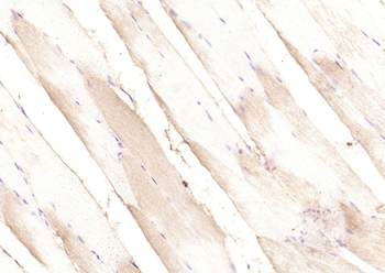
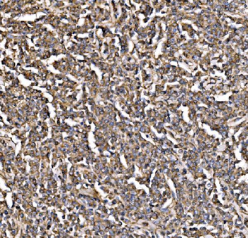
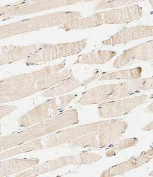
Paraformaldehyde-fixed, paraffin embedded (mouse skeletal muscle), Antigen retrieval by boiling in sodium citrate buffer (pH6.0) for 15 min, Block endogenous peroxidase by 3% hydrogen peroxide for 20 minutes, Blocking buffer (normal goat serum) at 37°C for 30 min, Antibody incubation with (PI 3 Kinase p85 alpha) Polyclonal Antibody, Unconjugated (orb11271) at 1:200 overnight at 4°C, followed by operating according to SP Kit (Rabbit) instructionsand DAB staining.
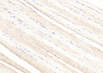
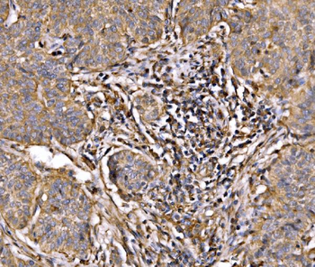
Paraformaldehyde-fixed, paraffin embedded (rat skeletal muscle), Antigen retrieval by boiling in sodium citrate buffer (pH6.0) for 15 min, Block endogenous peroxidase by 3% hydrogen peroxide for 20 minutes, Blocking buffer (normal goat serum) at 37°C for 30 min, Antibody incubation with (PI 3 Kinase p85 alpha) Polyclonal Antibody, Unconjugated (orb11271) at 1:200 overnight at 4°C, followed by operating according to SP Kit (Rabbit) instructionsand DAB staining.
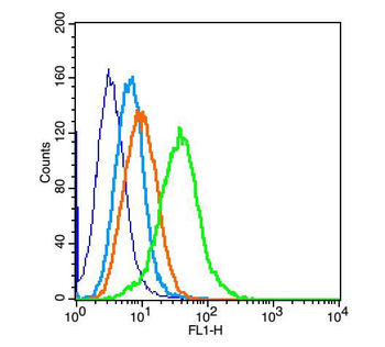
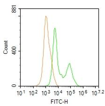
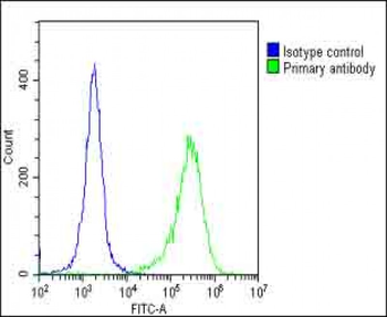
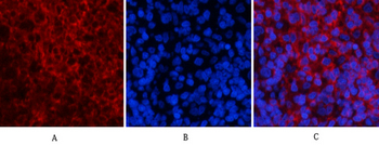
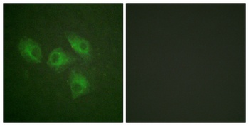
Positive control: H9C2 (2% Paraformaldehyde-fixed), Isotype Control Antibody, Antibody: Rabbit IgG, Dilution: 1 µg in 100 µl 1 X PBS containing 0.5% BSA, Secondary Antibody, Antibody: Goat anti-rabbit IgG-FITC, Dilution: 1:200 in 1 X PBS containing 0.5% BSA, Primary Antibody, orb10295, Dilution: 1 µg in 100 µl 1X PBS containing 0.5% BSA.
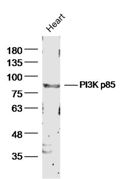
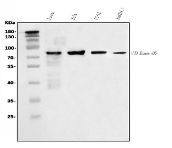
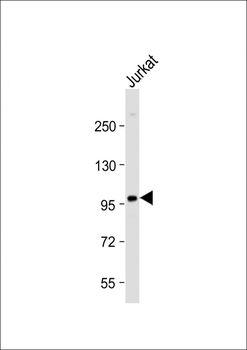
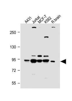
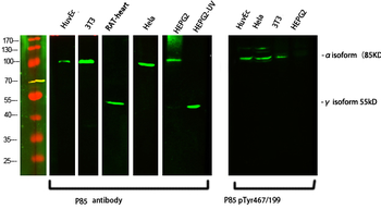
Sample: Heart (mouse) Lysate at 40 ug, Primary: Anti-PI3K p85 (orb11271) at 1/300 dilution, Secondary: IRDye800CW Goat Anti-Rabbit IgG at 1/20000 dilution, Predicted band size: 80kD, Observed band size: 85 kD.
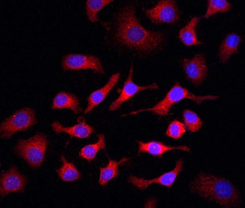
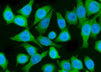
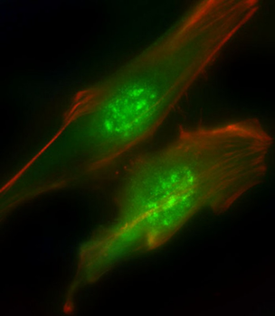
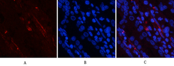
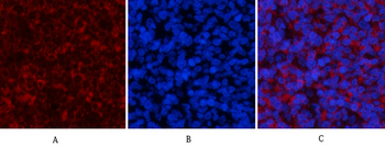
Tissue/Cell: HepG2 cell, 4% Paraformaldehyde-fixed, Triton X-100 at room temperature for 20 min, Blocking buffer (normal goat serum) at 37°C for 20 min, Antibody incubation with (PI3K p85) polyclonal Antibody, Unconjugated (orb11271) 1:100, 90 minutes at 37°C, followed by a FITC conjugated Goat Anti-Rabbit IgG antibody at 37°C for 90 minutes, DAPI (blue) was used to stain the cell nuclei.
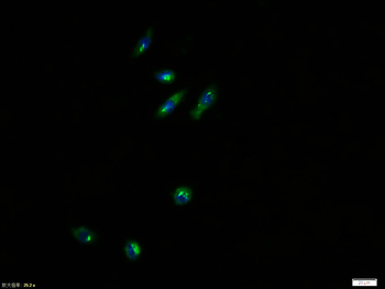
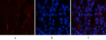
Tissue/Cell: NIH/3T3 cell, 4% Paraformaldehyde-fixed, Triton X-100 at room temperature for 20 min, Blocking buffer (normal goat serum) at 37°C for 20 min, Antibody incubation with (PI 3 Kinase p85 alpha) polyclonal Antibody, Unconjugated (orb11271) 1:100, 90 minutes at 37°C, followed by a FITC conjugated Goat Anti-Rabbit IgG antibody at 37°C for 90 minutes, DAPI (blue) was used to stain the cell nuclei.
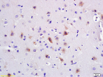
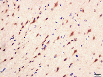
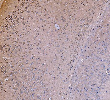
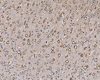
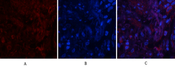
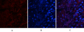
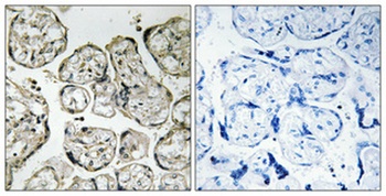
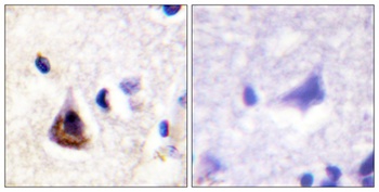
Tissue/Cell: rat brain tissue, 4% Paraformaldehyde-fixed and paraffin-embedded, Antigen retrieval: citrate buffer (0.01M, pH6.0), Boiling bathing for 15 min, Block endogenous peroxidase by 3% Hydrogen peroxide for 30 min, Blocking buffer (normal goat serum) at 37°C for 20 min, Incubation: Anti-PI3K/PI3 kinase p85 alpha subunit Polyclonal Antibody, Unconjugated (orb11271) 1:200, overnight at 4°C, followed by conjugation to the secondary antibody and DAB staining.
Quick Database Links
Gene Symbol
PIK3R1
UniProt
UniProt Details
− No UniProt data available
Documents Download
Datasheet
Product Information
Request a Document
Protocol Information
IHC-P
Immunohistochemistry Paraffin
IHC-Fr
Immunohistochemistry Frozen
IF
Immunofluorescence
Meng, Yingying et al. Oleic acid stimulates HC11 mammary epithelial cells proliferation and mammary gland development in peripubertal mice through activation of CD36-Ca2+ and PI3K/Akt signaling pathway Oncotarget, 9, 12982-12994 (2018)
PIK3R1 Rabbit Polyclonal Antibody (orb11271)

- 0.0
Based on 0 reviews
Participating in our Biorbyt product reviews program enables you to support fellow scientists by sharing your firsthand experience with our products.
Login to Submit a ReviewAvailable Sizes
Select a size below
Free Secondary Antibody (20 ul)0/0
Please add an antibody product to your cart first.