You have no items in your shopping cart.
Description
Research Area
Protein Biochemistry, Signal Transduction
Images & Validation
−Item 1 of 3
| Tested Applications | ELISA, ICC, IF, IP, WB |
|---|---|
| Dilution Range | WB (1:500), ICC/IF (1:60), ELISA (1:2000), IP (1:100) |
| Reactivity | All |
| Application Notes |
Key Properties
−| Host | Rabbit |
|---|---|
| Clonality | Polyclonal |
| Immunogen | Phosphothreonine conjugated to KLH |
| Target | Phosphothreonine |
| Purification | Peptide Affinity Purified |
| Conjugation | Unconjugated |
Storage & Handling
−| Storage | Maintain refrigerated at 2-8°C for up to 2 weeks. For long term storage store at -20°C in small aliquots to prevent freeze-thaw cycles. |
|---|---|
| Buffer/Preservatives | PBS, 50% glycerol, 0.01% sodium azide. Storage buffer changes when conjugated. |
| Concentration | 0.25 mg/ml |
| Expiration Date | 12 months from date of receipt. |
| Disclaimer | For research use only |
Alternative Names
−O-phospho-L-threonine, Phospho-threonine
Similar Products
−- Item 1 of 3
Phospho-Serine/Threonine Rabbit Polyclonal Antibody [orb158370]
ELISA, ICC, IF, IHC-Fr, IHC-P
Other
All
Rabbit
Polyclonal
Unconjugated
50 μl, 100 μl, 200 μl - Item 1 of 4
- Item 1 of 4
- Item 1 of 3
Phosphothreonine Antibody (Biotin) [orb396903]
ELISA, ICC, IF, IP, WB
All
Rabbit
Polyclonal
Biotin
400 μl - Item 1 of 3


Quality Guarantee
Explore bioreagents carefree to elevate your research. All our products are rigorously tested for performance. If a product does not perform as described on its datasheet, our scientific support team will provide expert troubleshooting, a prompt replacement, or a refund. For full details, please see our Terms & Conditions and Buying Guide. Contact us at [email protected].

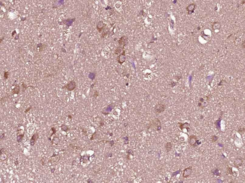
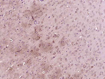
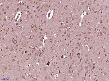
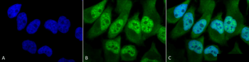
Immunocytochemistry/Immunofluorescence analysis using Rabbit Anti-Phosphothreonine Polyclonal Antibody. Tissue: Cervical cancer cell line (HeLa). Species: Human. Fixation: 2% Formaldehyde for 20 min at RT. Primary Antibody: Rabbit Anti-Phosphothreonine Polyclonal Antibody at 1:60 for 12 hours at 4°C. Secondary Antibody: FITC Goat Anti-Rabbit (green) at 1:200 for 2 hours at RT. Counterstain: DAPI (blue) nuclear stain at 1:40000 for 2 hours at RT. Localization: Cytoplasm. Nucleus. Magnification: 100x. (A) DAPI (blue) nuclear stain. (B) Anti-Phosphothreonine Antibody. (C) Composite.

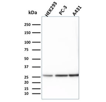
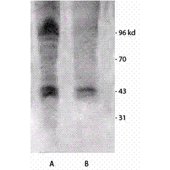
Western blot analysis of Mouse brain cell lysates showing detection of Phosphothreonine protein using Rabbit Anti-Phosphothreonine Polyclonal Antibody. Primary Antibody: Rabbit Anti-Phosphothreonine Polyclonal Antibody at 1:1000. Left: Treated with Vanadium, Right: Non-treated.

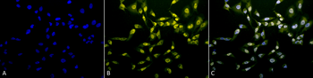
Immunocytochemistry/Immunofluorescence analysis using Rabbit Anti-Phosphothreonine Polyclonal Antibody. Tissue: Cervical cancer cell line (HeLa). Species: Human. Fixation: 2% Formaldehyde for 20 min at RT. Primary Antibody: Rabbit Anti-Phosphothreonine Polyclonal Antibody at 1:60 for 12 hours at 4°C. Secondary Antibody: R-PE Goat Anti-Rabbit (yellow) at 1:200 for 2 hours at RT. Counterstain: DAPI (blue) nuclear stain at 1:40000 for 2 hours at RT. Localization: Cytoplasm. Nucleus. Magnification: 20x. (A) DAPI (blue) nuclear stain. (B) Anti-Phosphothreonine Antibody. (C) Composite.
Quick Database Links
Gene Symbol
Phosphothreonine
Documents Download
Datasheet
Product Information
Request a Document
Protocol Information
WB
Western Blot (IB, immunoblot)
IF
Immunofluorescence
ICC
Immunocytochemistry
ELISA
Enzyme-linked Immunosorbent Assay (EIA)
IP
Immunoprecipitation
Phosphothreonine Antibody (orb613991)

- 0.0
Based on 0 reviews
Participating in our Biorbyt product reviews program enables you to support fellow scientists by sharing your firsthand experience with our products.
Login to Submit a ReviewAvailable Sizes
Select a size below
Free Secondary Antibody (20 ul)0/0
Please add an antibody product to your cart first.