You have no items in your shopping cart.
Featured
KO/KD
Validated
Validated
Description
Research Area
Cell Biology
Images & Validation
−Item 1 of 9
| Tested Applications | ELISA, IF, IHC-P, KO/KD Validated, WB |
|---|---|
| Reactivity | Human, Mouse, Rat |
| Predicted Reactivity | Bovine |
Key Properties
−| Antibody Type | Primary Antibody |
|---|---|
| Host | Rabbit |
| Clonality | Polyclonal |
| Isotype | IgG |
| Immunogen | Anti-PHAP I antibody (orb1239868) was raised against a peptide corresponding to 14 amino acids near the carboxy terminus of human PHAP I . The immunogen is located within the last 50 amino acids of PHAP I. |
| Target | ANP32A |
| Molecular Weight | Predicted: 29kDObserved: 29 kD |
| Purification | PHAP I Antibody is DEAE purified. |
| Conjugation | Unconjugated |
Storage & Handling
−| Storage | Maintain refrigerated at 2-8°C for up to 2 weeks. For long term storage store at -20°C in small aliquots to prevent freeze-thaw cycles. |
|---|---|
| Form/Appearance | Liquid |
| Buffer/Preservatives | PHAP I Antibody is supplied in PBS containing 0.02% sodium azide. |
| Concentration | 1 mg/mL |
| Expiration Date | 12 months from date of receipt. |
| Disclaimer | For research use only |
Alternative Names
−PHAP I Antibody: LANP, MAPM, PP32, HPPCn, PHAP1, PHAPI, I1PP2A, C15orf1, LANP, Acidic leucine-rich nuclear phosphoprotein 32 family member A, Acidic nuclear phosphoprotein pp32
Similar Products
−- Item 1 of 9
ANP32A Antibody [orb1239865]
ELISA, ICC, IF, KO/KD Validated, WB
Human, Mouse, Rat
Rabbit
Polyclonal
Unconjugated
0.02 mg, 0.1 mg - Item 1 of 5
PHAP1 Rabbit Polyclonal Antibody [orb100709]
IF, IHC-Fr, IHC-P, WB
Bovine, Canine, Equine, Porcine, Rabbit
Human, Mouse, Rat
Rabbit
Polyclonal
Unconjugated
50 μl, 100 μl, 200 μl - Item 1 of 3
ANP32A Antibody (C-term) [orb1927878]
IHC-P, WB
Bovine
Human
Rabbit
Polyclonal
Unconjugated
50 μl, 100 μl 
ANP32A Antibody [orb3162403]
ELISA, ICC, IHC, WB
Human, Mouse, Rat
Rabbit
Polyclonal
Unconjugated
50 μl, 100 μlANP32A Antibody [orb3162402]
ELISA, IHC, WB
Human, Mouse, Rat
Rabbit
Polyclonal
Unconjugated
50 μl, 100 μl


Quality Guarantee
Explore bioreagents carefree to elevate your research. All our products are rigorously tested for performance. If a product does not perform as described on its datasheet, our scientific support team will provide expert troubleshooting, a prompt replacement, or a refund. For full details, please see our Terms & Conditions and Buying Guide. Contact us at [email protected].
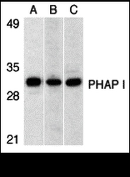
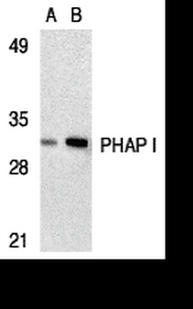
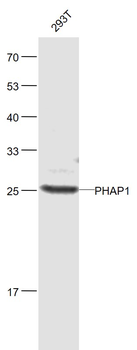
Western Blot Validation in (A) Human Raji Cells, (B) Mouse testis tissue lysate and (C) Rat testis tissue lysate. Loading: 15 µg of lysates per lane. Antibodies: PHAP I orb1239868 (1 µg/mL), 1h incubation at RT in 5% NFDM/TBST. Secondary: Goat anti-rabbit IgG HRP conjugate at 1:10000 dilution.

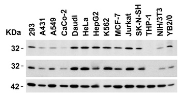
Independent Antibody Validation (IAV) via Protein Expression Profile in Cell Lines. Loading: 15 µg of lysates per lane. Antibodies: PHAP I orb1239865 (2 µg/mL), PHAP I orb1239868 (1 µg/mL), and beta-actin (1 µg/mL), 1h incubation at RT in 5% NFDM/TBST. Secondary: Goat anti-rabbit IgG HRP conjugate at 1:10000 dilution.
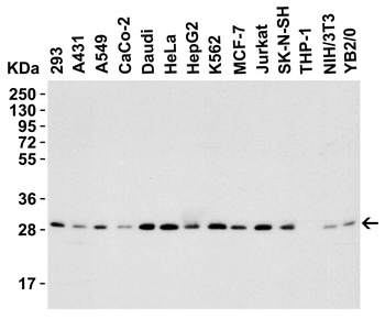
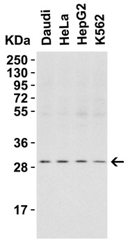
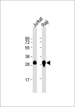
Western Blot Validation in Human, Mouse and Rat Cell Lines. Loading: 15 µg of lysates per lane. Antibodies: PHAP I orb1239868 (1 µg/mL), 1h incubation at RT in 5% NFDM/TBST. Secondary: Goat anti-rabbit IgG HRP conjugate at 1:10000 dilution.
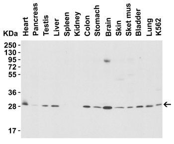
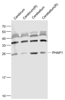
Western Blot Validation in Mouse Tissues. Loading: 15 µg of lysates per lane. Antibodies: PHAP I orb1239868 (1 µg/mL), 1h incubation at RT in 5% NFDM/TBST. Secondary: Goat anti-rabbit IgG HRP conjugate at 1:10000 dilution.
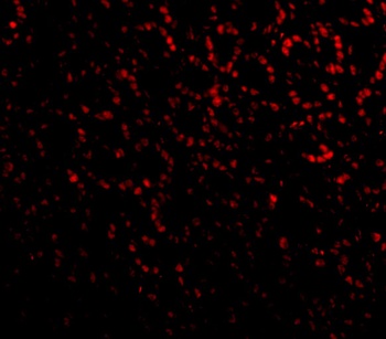
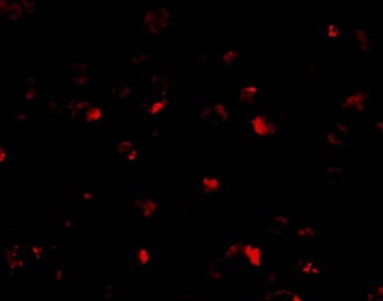
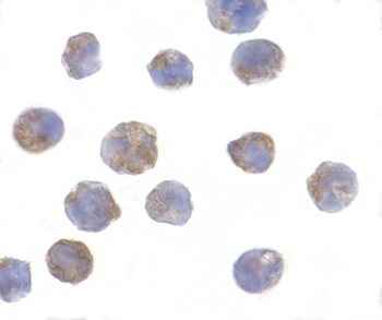
Immunofluorescence Validation of PHAP I in Mouse Small Intestine cells. Immunofluorescent analysis of 4% paraformaldehyde-fixed Mouse Small Intestine Cells labeling PHAP I with orb1239868 at 20 µg/mL, followed by goat anti-rabbit IgG secondary antibody at 1/500 dilution (red).
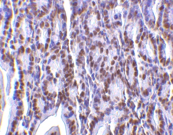
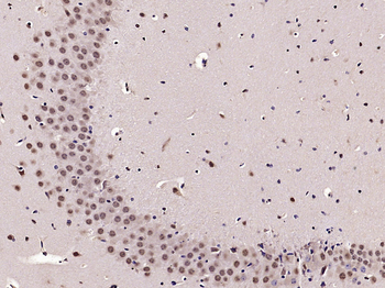
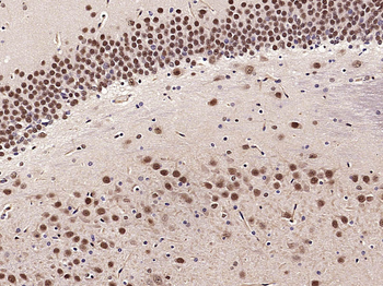
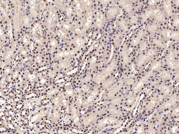
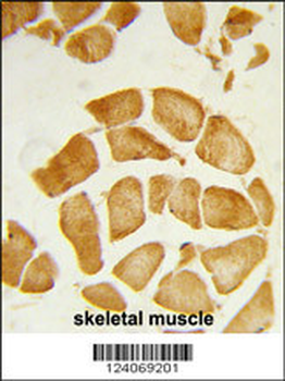
Immunohistochemistry Validation of PHAP I in Mouse Small Intestine Tissue. Immunohistochemical analysis of paraffin-embedded Mouse Small Intestine Tissue using anti-PHAP I antibody (orb1239868) at 2 µg/ml. Tissue was fixed with formaldehyde and blocked with 10% serum for 1 h at RT; antigen retrieval was by heat mediation with a citrate buffer (pH6). Samples were incubated with primary antibody overnight at 4°C. A goat anti-rabbit IgG H&L (HRP) at 1/250 was used as secondary. Counter stained with Hematoxylin.
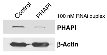
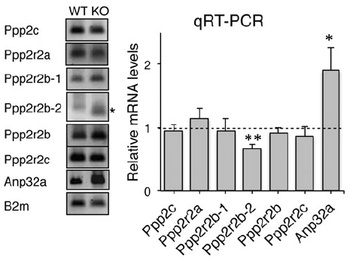
KD Validation of PHAPI in Human Breast Cancer Cells (Schafer et al., 2006). Human breast cancer cells (T47D cells) were transfected with control or PHAPI siRNA duplex. PHAPI was detected via Western Blot analysis by using the anti-PHAPI antibody. PHAPI expression was reduced after PHAPI siRNA knockdown.
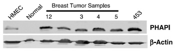
Increased Expression Validation of PHAPI in Patient Samples of BreastTumor Tissue (Schafer et al., 2006). PHAPI was overexpressed in all breast tumor samples of patients and human breast cancer cells (MDA-MB-453), but not in the normal breast tissue or human primary mammary epithelial cells (HMEC).
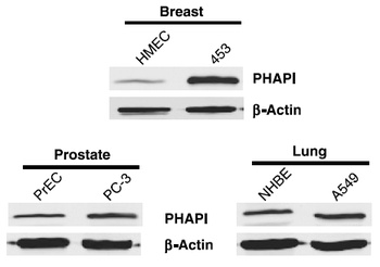
Overexpression of PHAPI in Breast Cancer Cells (Schafer et al., 2006). Western blot analysis with anti-PHAPI antibodies was performed for PHAPI in human cell lines from breast, prostate and lung. PHAPI was overexpressed in breast cancer cells when compared with normal cells (HMEC) whereas there were no significant differences in PHAPI expression in normal and cancer cells of either prostate or lung origin.
Documents Download
Datasheet
Product Information
Request a Document
Protocol Information
WB
Western Blot (IB, immunoblot)
IHC-P
Immunohistochemistry Paraffin
IF
Immunofluorescence
ELISA
Enzyme-linked Immunosorbent Assay (EIA)
ANP32A Antibody (orb1239868)

- 0.0
Based on 0 reviews
Participating in our Biorbyt product reviews program enables you to support fellow scientists by sharing your firsthand experience with our products.
Login to Submit a ReviewAvailable Sizes
Select a size below
Free Secondary Antibody (20 ul)0/0
Please add an antibody product to your cart first.