You have no items in your shopping cart.
Featured
Description
Research Area
Inflammation, Inflammatory Mediators, Oxidative Stress
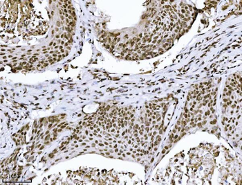
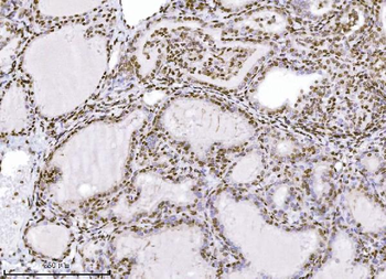
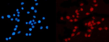
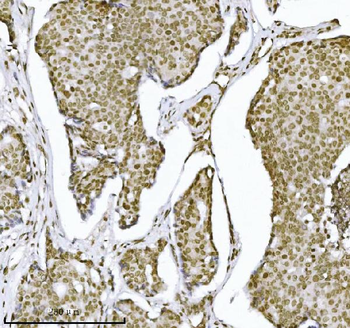
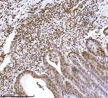
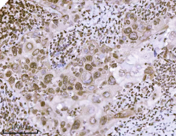
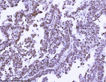
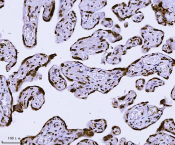
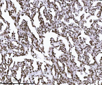
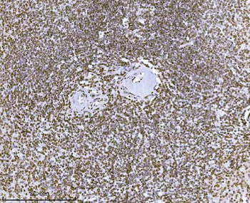
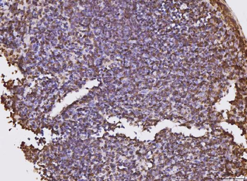
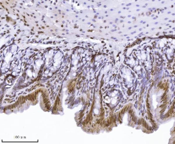
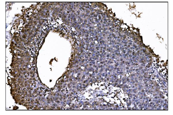
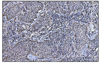
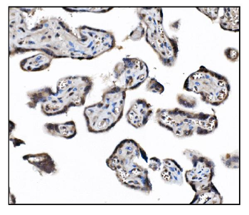
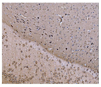
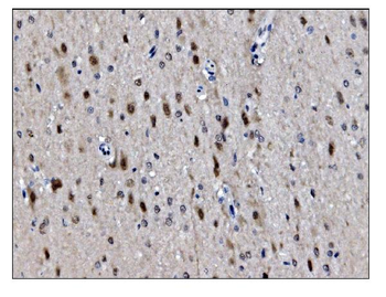
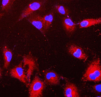
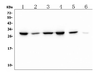
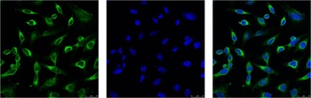
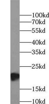
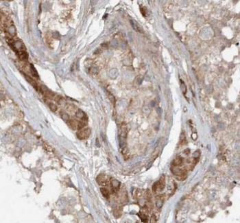
Images & Validation
−
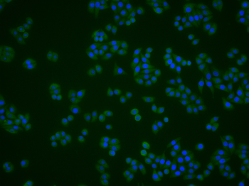
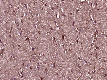
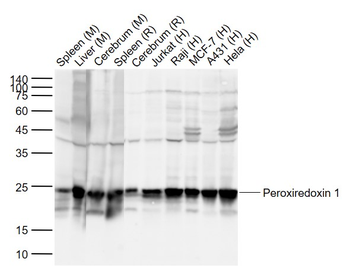
Item 1 of 4
| Tested Applications | FC, IF, IHC-Fr, IHC-P, WB |
|---|---|
| Dilution Range | WB=1:500-1000, IHC-P=1:100-500, IHC-F=1:100-500, IF=1:100-500, Flow-Cyt=0.5ug/Test |
| Reactivity | Human, Mouse, Rat |
| Predicted Reactivity | Mouse, Rat |
Related Conjugates & Formulations
−Key Properties
−| Antibody Type | Primary Antibody |
|---|---|
| Host | Mouse |
| Clonality | Monoclonal |
| Isotype | IgG |
| Clone No. | B32H1 |
| Immunogen | Recombinant human Peroxiredoxin 1 Protein |
| Target | PRDX1 |
| Molecular Weight | 23 kDa |
| Purification | Affinity purified by Protein G |
| Conjugation | Unconjugated |
Storage & Handling
−| Storage | Maintain refrigerated at 2-8°C for up to 2 weeks. For long term storage store at -20°C in small aliquots to prevent freeze-thaw cycles. |
|---|---|
| Form/Appearance | Liquid |
| Buffer/Preservatives | 0.01M TBS (pH7.4) with 1% rAlbumin, 0.02% Proclin300 and 50% Glycerol. |
| Concentration | 1mg/ml |
| Expiration Date | 12 months from date of receipt. |
| Disclaimer | For research use only |
Alternative Names
−PRX1; MSP23; NKEF-A; NKEFA; PAG; PAGA; PAGB; PRXI; TDPX2; OSF-3; OSF3; PrdxI; TDX2; TPxA; Hbp23; PRDX1_CRIGR; PRDX1; Thioredoxin peroxidase 2 (TPX-2); Thioredoxin-dependent peroxiredoxin 1; 1.11.1.24; PRDX1_HUMAN; Natural killer cell-enhancing factor A (NKEF-A); Proliferation-associated gene protein (PAG); Thioredoxin peroxidase 2; Thioredoxin-dependent peroxide reductase 2; PRDX1_MOUSE; Macrophage 23 kDa stress protein; Osteoblast-specific factor 3 (OSF-3); PRDX1_RAT; Heme-binding 23 kDa protein; peroxiredoxin 1
Similar Products
−- Item 1 of 14
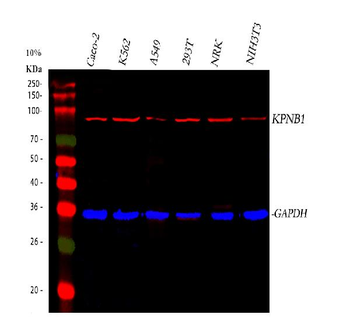
KPNB1 Mouse Monoclonal Antibody [orb1152394]
FC, ICC, IF, IHC, WB
Human, Mouse, Rat
Mouse
Monoclonal
Unconjugated
100 μg - Item 1 of 9
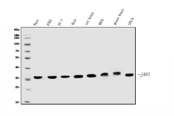
JAB1 Mouse Monoclonal Antibody [orb763099]
FC, ICC, IF, IHC, WB
Human, Mouse, Rat
Mouse
Monoclonal
Unconjugated
100 μg - Item 1 of 4
PRDX6 Mouse Monoclonal Antibody [orb623773]
FC, ICC, IF, IHC, WB
Human, Mouse, Rat
Mouse
Monoclonal
Unconjugated
100 μg - Item 1 of 2
Peroxiredoxin 1 Mouse mAb Antibody [orb763458]
IF, WB
Human, Mouse, Rat
Monoclonal
Unconjugated
50 μl, 100 μl - Item 1 of 2
PRDX2 Mouse Monoclonal Antibody [orb395421]
ELISA, IF, IHC, WB
Human, Mouse
Mouse
Monoclonal
Unconjugated
50 μg, 100 μg


Quality Guarantee
Explore bioreagents carefree to elevate your research. All our products are rigorously tested for performance. If a product does not perform as described on its datasheet, our scientific support team will provide expert troubleshooting, a prompt replacement, or a refund. For full details, please see our Terms & Conditions and Buying Guide. Contact us at [email protected].
Quick Database Links
Gene Symbol
PRDX1
UniProt
UniProt Details
− No UniProt data available
Protocol Information
WB
Western Blot (IB, immunoblot)
IHC-P
Immunohistochemistry Paraffin
IHC-Fr
Immunohistochemistry Frozen
FC
Flow Cytometry
IF
Immunofluorescence
Available Sizes
Select a size below
Free Secondary Antibody (20 ul)0/0
Please add an antibody product to your cart first.