You have no items in your shopping cart.
KO/KD
Validated
Validated
Description
Research Area
Signal Transduction
Images & Validation
−Item 1 of 5
| Tested Applications | IHC-P, KO/KD Validated, WB |
|---|---|
| Dilution Range | WB - 1:1000, IHC-P - 1:25 |
| Reactivity | Human, Mouse |
Key Properties
−| Host | Mouse |
|---|---|
| Clonality | Monoclonal |
| Isotype | IgG1,K |
| Clone No. | B302EV32X4X5 |
| Immunogen | This PDK2 monoclonal antibody is generated from mouse immunized with PDK2 recombinant protein. |
| Target | PDK2 |
| Molecular Weight | 46154 Da |
| Conjugation | Unconjugated |
Storage & Handling
−| Storage | Maintain refrigerated at 2-8°C for up to 2 weeks. For long term storage store at -20°C in small aliquots to prevent freeze-thaw cycles |
|---|---|
| Form/Appearance | Purified monoclonal antibody supplied in PBS with 0.09% (W/V) sodium azide. This antibody is purified through a protein G column, followed by dialysis against PBS. |
| Expiration Date | 12 months from date of receipt. |
| Disclaimer | For research use only |
Alternative Names
−[Pyruvate dehydrogenase (acetyl-transferring)] kinase isozyme 2, mitochondrial, Pyruvate dehydrogenase kinase isoform 2, PDH kinase 2, PDKII, PDK2, PDHK2
Similar Products
−- Item 1 of 1
Mouse Pyruvate Dehydrogenase Kinase Isozyme 2 (PDK2) ELISA Kit [orb781182]
Mouse
0.16-10 ng/mL
0.06 ng/mL
48 T, 96 T - Item 1 of 5
- Item 1 of 4
Mouse Pdk2 Antibody (N-term) [orb1936154]
IHC-P, WB
Rat
Human, Mouse
Rabbit
Polyclonal
Unconjugated
100 μl, 50 μl - Item 1 of 3
- Item 1 of 4
PDK2 Rabbit Polyclonal Antibody [orb137873]
FC, ICC, IF, IP, WB
Human, Mouse, Rat
Rabbit
Polyclonal
Unconjugated
100 μg


Quality Guarantee
Explore bioreagents carefree to elevate your research. All our products are rigorously tested for performance. If a product does not perform as described on its datasheet, our scientific support team will provide expert troubleshooting, a prompt replacement, or a refund. For full details, please see our Terms & Conditions and Buying Guide. Contact us at [email protected].
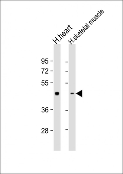
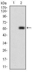
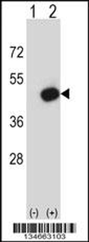
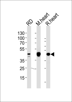
All lanes: Anti- at 1:1000 dilution. Lane 1: human heart lysate. Lane 2: human skeletal muscle lysate.Lysates/proteins at 20 µg per lane. Secondary Goat Anti-mouse IgG, (H+L), Peroxidase conjugated at 1/10000 dilution. Predicted band size: 46 kDa. Blocking/Dilution buffer: 5% NFDM/TBST.
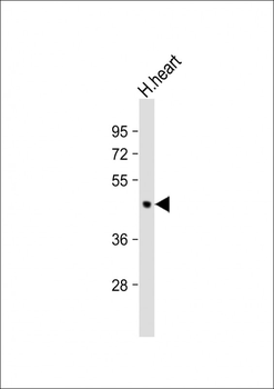
Anti-PDK2 Antibody at 1:1000 dilution + human heart lysate. Lysates/proteins at 20 µg per lane. Secondary Goat Anti-mouse IgG, (H+L), Peroxidase conjugated at 1/10000 dilution. Predicted band size: 46 kDa. Blocking/Dilution buffer: 5% NFDM/TBST.
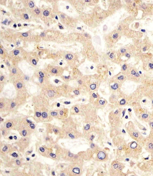
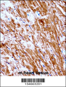
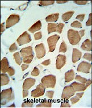
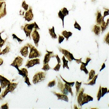
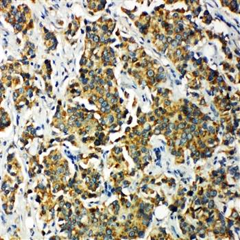
Staining PDK2 in human liver sections by Immunohistochemistry (IHC-P - paraformaldehyde-fixed, paraffin-embedded sections). Tissue was fixed with formaldehyde and blocked with 3% BSA for 0.5 hour at room temperature; antigen retrieval was by heat mediation with a citrate buffer (pH6). Samples were incubated with primary antibody (1/25) for 1 hours at 37°C. A undiluted biotinylated goat polyvalent antibody was used as the secondary Antibody.
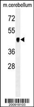
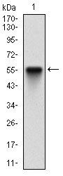
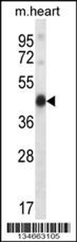
PDK2 Antibody western blot analysis in mouse cerebellum tissue lysates (35 μg/lane). This demonstrates the PDK2 antibody detected the PDK2 protein (arrow).
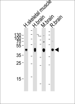
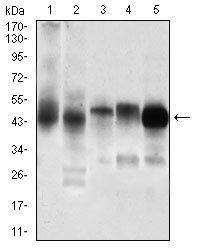
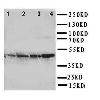
Western blot analysis of lysates from human skeletal muscle, human brain, mouse brain, rat brain tissue (from left to right), using PDK2 Antibody. diluted at 1:2000 at each lane. A goat anti-mouse IgG H&L (HRP) at 1:3000 dilution was used as the secondary Antibody. Lysates at 20 μg per lane.
Quick Database Links
UniProt Details
− No UniProt data available
NCBI Reference Sequences
−Associated Accession Numbers
Curated reference sequences for the gene transcript and protein product| Protein | NP_002602.2, NP_001186827.1, NP_001186828.1 |
|---|
Documents Download
Datasheet
Product Information
Request a Document
Protocol Information
WB
Western Blot (IB, immunoblot)
IHC-P
Immunohistochemistry Paraffin
PDK2 Antibody (orb1939394)

- 0.0
Based on 0 reviews
Participating in our Biorbyt product reviews program enables you to support fellow scientists by sharing your firsthand experience with our products.
Login to Submit a ReviewAvailable Sizes
Select a size below
Choose Conjugation or Carrier Free Version
Free Secondary Antibody (20 ul)0/0
Please add an antibody product to your cart first.