You have no items in your shopping cart.
PDI Antibody
SKU: orb1822442
Description
Research Area
Cell Biology, Disease Biomarkers, Protein Biochemistry, Signal Transduction
Images & Validation
−Item 1 of 3
| Tested Applications | ICC, IF, IHC, IP, WB |
|---|---|
| Dilution Range | WB (1:1000), ICC/IF (1:100); optimal dilutions for assays should be determined by the user. |
| Reactivity | Bovine, Canine, Frog, Guinea pig, Hamster, Human, Invertebrate, Mouse, Mussel, Porcine, Rat, Sheep |
| Application Notes |
Key Properties
−| Host | Rabbit |
|---|---|
| Clonality | Polyclonal |
| Immunogen | AA499-509 of Rat PDI |
| Target | PDI |
| Molecular Weight | 58kDa |
| Purification | Protein A purified |
| Conjugation | Unconjugated |
Storage & Handling
−| Storage | Maintain refrigerated at 2-8°C for up to 2 weeks. For long term storage store at -20°C in small aliquots to prevent freeze-thaw cycles. |
|---|---|
| Buffer/Preservatives | PBS pH 7.4, 50% glycerol, 0.09% sodium azide Storage buffer changes when conjugated |
| Concentration | 1 mg/ml |
| Expiration Date | 12 months from date of receipt. |
| Disclaimer | For research use only |
Alternative Names
−Thioredoxin domain containing 5, ERP46, UNQ364, MGC3178, FLJ21353, FLJ90810, thioredoxin related protein, endothelial protein disulphide isomeras
Similar Products
−- Item 1 of 10
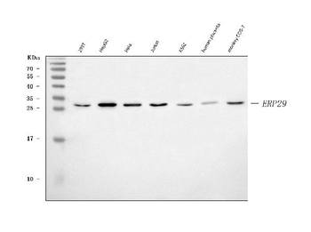
ERP29 Rabbit Polyclonal Antibody [orb1291744]
ELISA, FC, ICC, IF, IHC, WB
Human, Monkey, Mouse, Rat
Rabbit
Polyclonal
Unconjugated
100 μg - Item 1 of 6
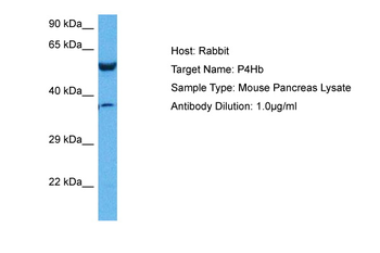
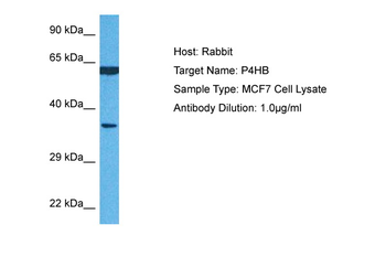
P4HB Rabbit Polyclonal Antibody [orb580304]
IHC, WB
Bovine, Canine, Equine, Guinea pig, Rabbit, Rat
Human, Mouse
Rabbit
Polyclonal
Unconjugated
100 μl - Item 1 of 1
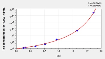
Human Protein Disulfide Isomerase A2 (PDIA2) ELISA Kit [orb775887]
Human
0.32-20 ng/mL
0.111 ng/mL
96 T, 48 T - Item 1 of 1
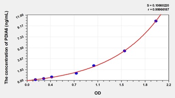
Human Protein Disulfide Isomerase A6 (PDIA6) ELISA Kit [orb777486]
Human
0.16-10 ng/mL
0.05 ng/mL
96 T, 48 T - Item 1 of 1
Human Protein Disulfide Isomerase A4 (PDIA4) ELISA Kit [orb778189]
Human
1.57-100 ng/mL
0.61 ng/mL
96 T, 48 T


Quality Guarantee
Explore bioreagents carefree to elevate your research. All our products are rigorously tested for performance. If a product does not perform as described on its datasheet, our scientific support team will provide expert troubleshooting, a prompt replacement, or a refund. For full details, please see our Terms & Conditions and Buying Guide. Contact us at [email protected].
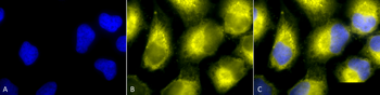
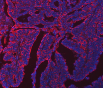
Immunocytochemistry/Immunofluorescence analysis using Rabbit Anti-PDI Polyclonal Antibody. Tissue: Cervical cancer cell line (HeLa). Species: Human. Fixation: 2% Formaldehyde for 20 min at RT. Primary Antibody: Rabbit Anti-PDI Polyclonal Antibody at 1:100 for 12 hours at 4°C. Secondary Antibody: R-PE Goat Anti-Rabbit (yellow) at 1:200 for 2 hours at RT. Counterstain: DAPI (blue) nuclear stain at 1:40000 for 2 hours at RT. Localization: Endoplasmic reticulum lumen. Melanosome. Magnification: 100x. (A) DAPI (blue) nuclear stain. (B) Anti-PDI Antibody. (C) Composite.
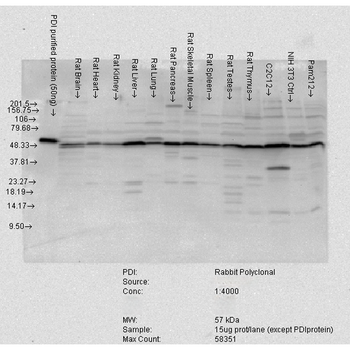
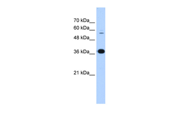
Western blot analysis of Rat tissue mix showing detection of PDI protein using Rabbit Anti-PDI Polyclonal Antibody. Load: 15 μgprotein. Block: 1.5% BSA. Primary Antibody: Rabbit Anti-PDI Polyclonal Antibody at 1:4000 for 2 hours at RT. Secondary Antibody: Donkey Anti-Rabbit IgG: HRP for 1 hour at RT.
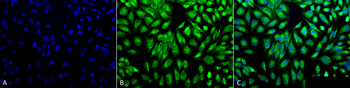
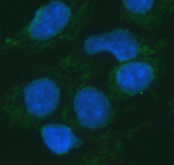
Immunocytochemistry/Immunofluorescence analysis using Rabbit Anti-PDI Polyclonal Antibody. Tissue: Cervical cancer cell line (HeLa). Species: Human. Fixation: 2% Formaldehyde for 20 min at RT. Primary Antibody: Rabbit Anti-PDI Polyclonal Antibody at 1:100 for 12 hours at 4°C. Secondary Antibody: FITC Goat Anti-Rabbit (green) at 1:200 for 2 hours at RT. Counterstain: DAPI (blue) nuclear stain at 1:40000 for 2 hours at RT. Localization: Endoplasmic reticulum lumen. Melanosome. Magnification: 20x. (A) DAPI (blue) nuclear stain. (B) Anti-PDI Antibody. (C) Composite.
Quick Database Links
UniProt Details
− No UniProt data available
NCBI Gene Details
− No NCBI Gene data available
NCBI Reference Sequences
−Associated Accession Numbers
Curated reference sequences for the gene transcript and protein product| Protein | NP_001099245.2 |
|---|
Documents Download
Datasheet
Product Information
Request a Document
Protocol Information
WB
Western Blot (IB, immunoblot)
IHC
Immunohistochemistry
IF
Immunofluorescence
ICC
Immunocytochemistry
IP
Immunoprecipitation
PDI Antibody (orb1822442)

- 0.0
Based on 0 reviews
Participating in our Biorbyt product reviews program enables you to support fellow scientists by sharing your firsthand experience with our products.
Login to Submit a ReviewAvailable Sizes
Select a size below