You have no items in your shopping cart.
KO/KD
Validated
Validated
Description
Research Area
Cell Biology
Images & Validation
−Item 1 of 8
| Tested Applications | 2D-PAGE, IHC, KO/KD Validated, WB |
|---|---|
| Dilution Range | IHC: 1:100, WB: 1:1000 |
| Reactivity | Human |
| Application Notes |
Key Properties
−| Antibody Type | Primary Antibody |
|---|---|
| Host | Rabbit |
| Clonality | Polyclonal |
| Isotype | IgG |
| Immunogen | PARP1 (N-term ZF1) purified antibody was prepared from whole rabbit serum produced by repeated immunizations with n-terminus region of human PARP1 zinc finger domain recombinant protein. |
| Target | PARP1 |
| Purity | PARP1 (N-term ZF1) was purified from monospecific antiserum by immunoaffinity chromatography using protein A coupled to agarose beads. This antibody is specific for human PARP1 protein. No cross reactivity detected towards other PARP members when using siRNAs against 18 PARP family members. Cross-reactivity with PARP1 from other sources has not been determined. |
| Conjugation | Unconjugated |
Storage & Handling
−| Storage | Store vial at -20° C prior to opening. Aliquot contents and freeze at -20° C or below for extended storage. Avoid cycles of freezing and thawing. Centrifuge product if not completely clear after standing at room temperature. This product is stable for several weeks at 4° C as an undiluted liquid. Dilute only prior to immediate use. |
|---|---|
| Form/Appearance | Liquid (sterile filtered) |
| Buffer/Preservatives | Preservative: 0.01% (w/v) Sodium Azide. Stabilizer: None; Buffer: 0.02 M Potassium Phosphate, 0.15 M Sodium Chloride, pH 7.2 |
| Concentration | 1.0 mg/ml |
| Expiration Date | 12 months from date of receipt. |
| Dry Ice Shipping | Please note: This product requires shipment on dry ice. A dry ice surcharge will apply. |
| Disclaimer | For research use only |
Alternative Names
−rabbit anti-PARP1 Antibody, Poly [ADP-ribose] polymerase 1, ADP-ribosyltransferase diphtheria toxin-like 1, ARTD1, NAD(+) ADP-ribosyltransferase 1, ADPRT 1, PPOL
Similar Products
−- Item 1 of 10
PARP/PARP1 Mouse Monoclonal Antibody [orb738400]
FC, ICC, IF, IHC, WB
Human, Mouse, Rat
Mouse
Monoclonal
Unconjugated
100 μg - Item 1 of 7
PARP/PARP1 Mouse Monoclonal Antibody [orb738402]
FC, ICC, IF, IHC, WB
Human, Mouse, Rat
Mouse
Monoclonal
Unconjugated
100 μg - Item 1 of 7
PARP/PARP1 Rabbit Polyclonal Antibody [orb234355]
FC, ICC, IF, IHC, WB
Human, Mouse, Rat
Rabbit
Polyclonal
Unconjugated
100 μg - Item 1 of 8
- Item 1 of 7
PARP1 Rabbit Polyclonal Antibody [orb704256]
FC, ICC, IF, IHC-Fr, IHC-P, KO/KD Validated, WB
Mouse, Rat
Human, Mouse
Rabbit
Polyclonal
Unconjugated
50 μl, 100 μl


Quality Guarantee
Explore bioreagents carefree to elevate your research. All our products are rigorously tested for performance. If a product does not perform as described on its datasheet, our scientific support team will provide expert troubleshooting, a prompt replacement, or a refund. For full details, please see our Terms & Conditions and Buying Guide. Contact us at [email protected].
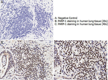
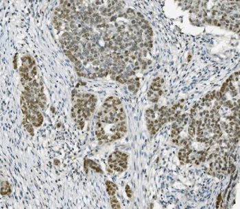
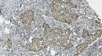
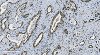
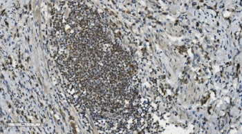
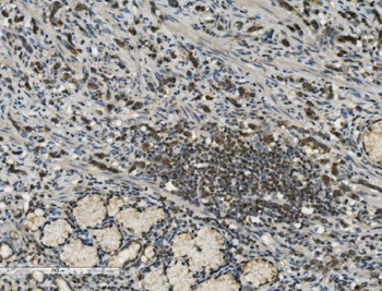
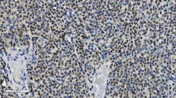
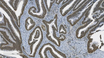
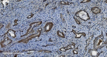
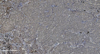
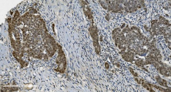
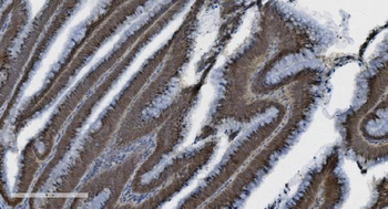
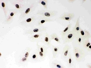
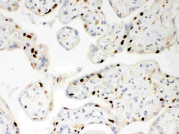
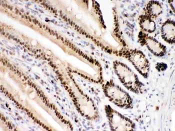
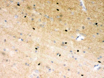
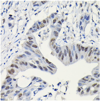
Immunohistochemistry with anti-PARP-1 antibody showing nuclear positivity in human lung tissue at 20x and 40x (B & C). Staining was performed on Leica Bond system using the standard protocol. Formalin fixed/paraffin embedded tissue sections were subjected to antigen retrieval and then incubated with rabbit anti-PARP-1 antibody orb420276 at 1:100 dilution for 60 minutes. Biotinylated Anti-rabbit secondary antibody was used at 1:200 dilution to detect primary antibody. The reaction was developed using streptavidin-HRP conjugated compact polymer system and visualized with chromogen substrate, 3'3-diamino-benzidine substrate (DAB). The sections were then counterstained with hematoxylin to detect cell nuclei.
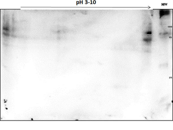
OVCAR-8 Wild Type Lysate was separated on 2D SDS-PAGE and blotted on PVDF to analyze immunocoverage of PARP1 antibody specific for the zinc finger 1 domain of PARP1. Primary Antibody: Anti-PARP1 (n-term) antibody 1:200 overnight at 4°C. Secondary Antibody: Goat anti-rabbit Peroxidase (orb347654) at 1:2000 at RT for 30 min. Blocking Buffer: BlockOut (p/n orb348644) for 30 min at RT. Predicted/observed: ~110 kDa and pI 9.7.

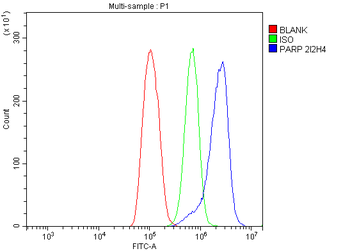
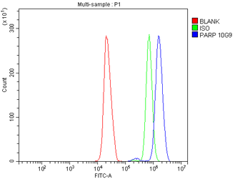
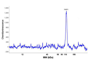
Peggy Sue™ Size Separation Electropherogram of OVCAR-8 lysates in no-salt buffer and detected with Anti-PARP1 (N-term ZF1). UV immobilization time: 250 seconds. Protein concentration: 577 µg/ml; 120 s UV immobilization. Primary antibody concentration: 20 µg/ml. Primary antibody incubation time: 180 min. Exposure time: multi-image analysis exposure. Predicted/observed: ~116 kDa.
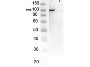
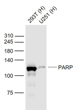
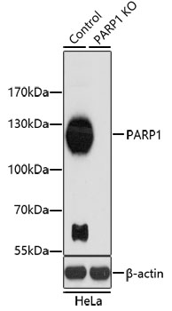
Western Blot of endogenous PARP1 with Rabbit Anti-PARP1 (N-term ZF1) Antibody. Lane 1: OVCAR8 Wild Type lysate. Lane 2: OVCAR8 PARP1 KO lysate. Load: 5 µg per lane. Primary antibody: PARP1 (N-term ZF1) antibody at 1 µg/ml for overnight at 4°C. Secondary antibody: HRP Gt-a-Rb IgG secondary antibody (p/n orb347654) at 1:40000 for 30 min at RT. Block: orb348637 overnight at 4°C. Predicted/Observed size: 113 kDa for endogenous PARP1. Other band(s): none.

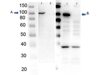
Western Blot of endogenous PARP1 with Rabbit Anti-PARP1 Antibodies. Lane 1: OVCAR8 Wild Type lysate. Lane 2: OVCAR8 PARP1 KO lysate. Load 5 µg per lane. Primary Antibody: Blot A: Anti-PARP1- n term (p/n orb420276); Blot B: Anti-PARP1- internal (p/n orb344711) at 1 µg/ml for overnight at 4°C. Secondary antibody: HRP Gt-a-Rb IgG secondary antibody (p/n orb347654) at 1:40000 for 30 min at RT. Block: orb348637 overnight at 4°C. Predicted/Observed size: 113 kDa for endogenous PARP1. Other band(s): nonspecific ~ 40 kDa in PARP1-AD only.

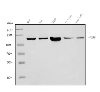
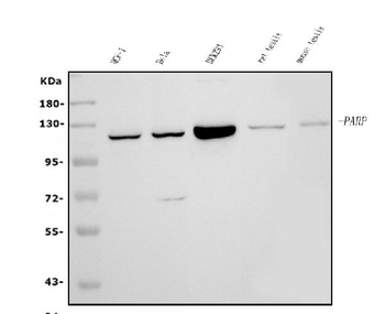
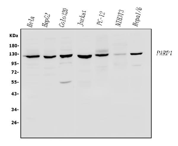
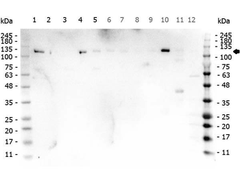
Western Blot of Rabbit anti-PARP1 antibody. Marker: Opal Pre-stained ladder. Lane 1: HEK293 lysate (p/n orb348669). Lane 2: HeLa Lysate (p/n orb348668). Lane 3: MCF-7 Lysate (p/n orb348664). Lane 4: Jurkat Lysate. Lane 5: A431 Lysate (p/n orb348665). Lane 6: A549 Lysate (p/n orb348675). Lane 7: LNCap Lysate (p/n orb348694). Lane 8: MOLT-4 Lysate (p/n orb348696). Lane 9: Ramos Lysate. Lane 10: Raji Lysate (p/n orb348672). Lane 11: A-172 Lysate (p/n orb348708). Lane 12: NIH/3T3 Lysate (p/n orb348714). Load: 35 µg per lane. Primary antibody: PARP1 antibody at 1 ug/ml overnight at 4C. Secondary antibody: Peroxidase rabbit secondary antibody (p/n orb347654) at 1:30000 for 60 min at RT. Blocking Buffer: 1% Casein-TTBS for 30 min at RT. Predicted/Observed size: 113 kDa for PARP1.

Western Blot of Rabbit anti-PARP1 N-term Antibody. Lane 1: Opal Pre-stained ladder. Lane 2: OVCAR-8 Wild Type. Lane 3: PARP1-KO. Lane 4: PARP2-KO. Lane 5: PARP3-KO. Lane 6: PARP4-KO Lane 7: PARP5a-KO. Lane 8: PARP5b-KO. Lane 9: PARP6-KO. Lane 10: PARP7-KO. Lane 11: PARP8-KO. Lane 12: PARP9-KO. Lane 13: PARP10-KO. Lane 14: PARP12-KO. Lane 15: PARP13-KO. Lane 16: PARP14-KO. Lane 17: PARP16-KO. Load: 5.0 µg per lane. Primary antibody: PARP1 n-term antibody at 1 ug/ml overnight at 4°C. Secondary antibody: Goat anti-rabbit Peroxidase secondary antibody (p/n orb347654) at 1:40000 for 30 min at RT. Blocking Buffer: orb348644 for 30 min at RT. Predicted/Observed size: ~113 kDa for PARP1.

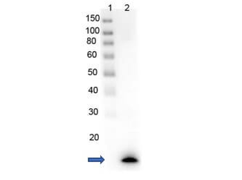
Western Blot of recombinant PARP1 with rabbit anti-PARP1 (N-term ZF1) antibody. Lane 1: PARP1-Zinc Finger domain recombinant protein. Load: 0.05 µg per lane. Primary antibody: PARP1 (N-term ZF1) antibody at 1 µg/ml for overnight at 4°C. Secondary antibody: HRP Gt-a-rabbit secondary antibody (p/n orb347654) at 1:40000 for 30 min at RT. Block: orb348637 overnight at 4°C. Predicted/Observed size: 13 kDa for rPARP1 (N-term ZF1). Other band(s): none.
Quick Database Links
Gene Symbol
PARP1
UniProt
UniProt Details
− No UniProt data available
Documents Download
Datasheet
Product Information
Request a Document
Protocol Information
WB
Western Blot (IB, immunoblot)
IHC
Immunohistochemistry
PARP1 Antibody (orb420276)

- 0.0
Based on 0 reviews
Participating in our Biorbyt product reviews program enables you to support fellow scientists by sharing your firsthand experience with our products.
Login to Submit a ReviewAvailable Sizes
Select a size below
Choose Conjugation or Carrier Free Version
Free Secondary Antibody (20 ul)0/0
Please add an antibody product to your cart first.