You have no items in your shopping cart.
Description
Research Area
Cell Biology, Immunology & Inflammation, Neuroscience, Signal Transduction
Images & Validation
−Item 1 of 4
| Tested Applications | IHC-P, WB |
|---|---|
| Dilution Range | WB - 1:1000, IHC-P - 1:100 |
| Reactivity | Human |
Key Properties
−| Antibody Type | Primary Antibody |
|---|---|
| Host | Rabbit |
| Clonality | Polyclonal |
| Isotype | Rabbit IgG |
| Immunogen | This PAK5 antibody is generated from rabbits immunized with a KLH conjugated synthetic peptide between 168-198 amino acids from human PAK5. Antigen Region: 168-198 aa. |
| Target | PAK5 (HGNC:15916) |
| Molecular Weight | 80745 Da |
| Conjugation | Unconjugated |
Storage & Handling
−| Storage | Maintain refrigerated at 2-8°C for up to 2 weeks. For long term storage store at -20°C in small aliquots to prevent freeze-thaw cycles |
|---|---|
| Form/Appearance | Purified polyclonal antibody supplied in PBS with 0.09% (W/V) sodium azide. This antibody is purified through a protein A column, followed by peptide affinity purification. |
| Expiration Date | 12 months from date of receipt. |
| Disclaimer | For research use only |
Alternative Names
−Serine/threonine-protein kinase PAK 7, p21-activated kinase 5, PAK-5, p21-activated kinase 7, PAK-7, PAK7, KIAA1264, PAK5
Similar Products
−- Item 1 of 9
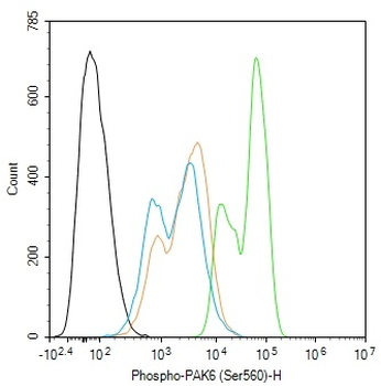
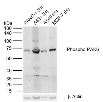
Phospho-PAK6 (Ser560) Rabbit Polyclonal Antibody [orb6634]
FC, ICC, IF, IHC-Fr, IHC-P, WB
Bovine, Canine, Equine, Gallus, Porcine, Rabbit, Sheep
Human, Mouse, Rat
Rabbit
Polyclonal
Unconjugated
100 μl, 50 μl, 200 μl - Item 1 of 3
Mouse Pak7 Antibody (N-term) [orb32169]
IHC-P, WB
Rat
Human, Mouse
Rabbit
Polyclonal
Unconjugated
50 μl, 100 μl - Item 1 of 3
- Item 1 of 1
PAK6 Rabbit Polyclonal Antibody [orb2951864]
ELISA, IHC, WB
Human
Rabbit
Polyclonal
Unconjugated
50 μg, 100 μg - Item 1 of 1


Quality Guarantee
Explore bioreagents carefree to elevate your research. All our products are rigorously tested for performance. If a product does not perform as described on its datasheet, our scientific support team will provide expert troubleshooting, a prompt replacement, or a refund. For full details, please see our Terms & Conditions and Buying Guide. Contact us at [email protected].

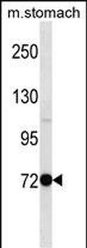
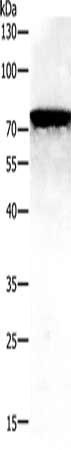
Anti-PAK5 Antibody at 1:2000 dilution + Human brain lysate. Lysates/proteins at 20 µg per lane. Secondary Goat Anti-Rabbit IgG, (H+L), Peroxidase conjugated at 1/10000 dilution. Predicted band size: 81 kDa. Blocking/Dilution buffer: 5% NFDM/TBST.
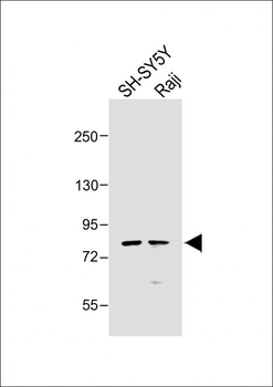
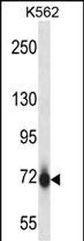
All lanes: Anti-PAK5 Antibody at 1:1000 dilution. Lane 1: SH-SY5Y whole cell lysate. Lane 2: Raji whole cell lysate. Lysates/proteins at 20 ug per lane. Secondary Goat Anti-Rabbit IgG, (H+L), Peroxidase conjugated at 1/10000 dilution. Predicted band size: 81 kDa. Blocking/Dilution buffer: 5% NFDM/TBST.
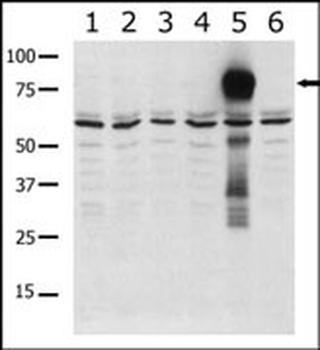
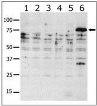
Western blot analysis of anti-PAK5 Pab in lysates from transiently transfected COS7 cells. Lane 1: negative control, Lane 2: PAK1-expressing cells, Lane 3: PAK2-expressing cells, Lane 4: PAK4-expressing cells, Lane 5: PAK5-expressing cells, and Lane 6: PAK6-expressing cells. PAK5 (arrow) was detected using purified Pab.
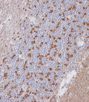
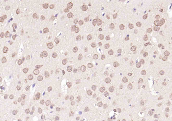
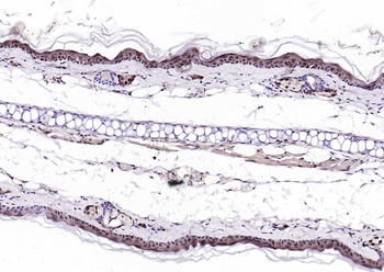
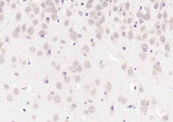
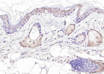
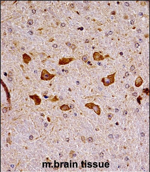
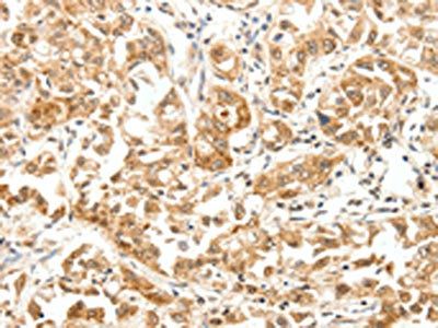
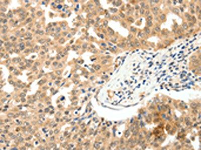
Immunohistochemical analysis on paraffin-embedded Human cerebellum tissue. Tissue was fixed with formaldehyde at room temperature. Heat induced epitope retrieval was performed by EDTA buffer (pH9.0). Samples were incubated with primary antibody (1:100) for 1 hour at room temperature. Undiluted CRF Anti-Polyvalent HRP Polymer antibody was used as the secondary antibody.
Quick Database Links
UniProt Details
− No UniProt data available
NCBI Reference Sequences
−Associated Accession Numbers
Curated reference sequences for the gene transcript and protein product| Protein | NP_817127.1, NP_065074.1 |
|---|
Documents Download
Datasheet
Product Information
Request a Document
Protocol Information
WB
Western Blot (IB, immunoblot)
IHC-P
Immunohistochemistry Paraffin
PAK5 Antibody (orb1928757)

- 0.0
Based on 0 reviews
Participating in our Biorbyt product reviews program enables you to support fellow scientists by sharing your firsthand experience with our products.
Login to Submit a ReviewAvailable Sizes
Select a size below
Choose Conjugation or Carrier Free Version
Free Secondary Antibody (20 ul)0/0
Please add an antibody product to your cart first.