You have no items in your shopping cart.
Description
Images & Validation
−Item 1 of 5
| Tested Applications | FC, ICC, IF, IHC |
|---|---|
| Reactivity | Human |
| Application Notes |
Key Properties
−| Antibody Type | Primary Antibody |
|---|---|
| Host | Mouse |
| Clonality | Monoclonal |
| Isotype | IgG1, kappa |
| Clone No. | NM95 |
| Immunogen | Nuclei of myeloid leukemia biopsy cells were used as the immunogen. |
| Purification | Protein G affinity chromatography |
| Conjugation | Unconjugated |
Storage & Handling
−| Storage | Maintain refrigerated at 2-8°C for up to 2 weeks. For long term storage store at -20°C in small aliquots to prevent freeze-thaw cycles. |
|---|---|
| Form/Appearance | Liquid |
| Buffer/Preservatives | PBS with 0.1 mg/ml rAlbumin and 0.05% sodium azide |
| Concentration | 0.2 mg/mL |
| Expiration Date | 12 months from date of receipt. |
| Disclaimer | For research use only |
Similar Products
−- Item 1 of 7
Nucleoli Marker Antibody / Human Nucleolar Antigen [orb248680]
FACS, ICC, IF, IHC-P
Human
Mouse
Monoclonal
Unconjugated
100 μg, 20 μg - Item 1 of 7
Nucleoli Marker Antibody / Human Nucleolar Antigen [orb2638091]
FACS, ICC, IF, IHC-P
Human
Mouse
Monoclonal
Unconjugated
100 μg - Item 1 of 4
Nucleoli Marker Antibody / Human Nucleolar Antigen [orb2638090]
IHC-P
Human
Mouse
Monoclonal
Unconjugated
7 ml 


Quality Guarantee
Explore bioreagents carefree to elevate your research. All our products are rigorously tested for performance. If a product does not perform as described on its datasheet, our scientific support team will provide expert troubleshooting, a prompt replacement, or a refund. For full details, please see our Terms & Conditions and Buying Guide. Contact us at [email protected].
![Nucleoli Marker Antibody [NM95]](/images/pub/media/catalog/product/NewWebsite/15/orb1252651_1.jpg)
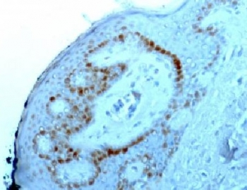
IHC staining of FFPE human skin with Nucleoli marker antibody.
![Nucleoli Marker Antibody [NM95]](/images/pub/media/catalog/product/NewWebsite/15/orb1252651_2.jpg)
IHC staining of FFPE human tonsil tissue with Nucleoli marker antibody.
![Nucleoli Marker Antibody [NM95]](/images/pub/media/catalog/product/NewWebsite/15/orb1252651_3.jpg)
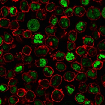
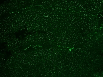
Immunofluorescent testing of colon carcinoma with Alexa Fluor 488 conjugated Nucleoli marker antibody.
![Nucleoli Marker Antibody [NM95]](/images/pub/media/catalog/product/NewWebsite/15/orb1252651_4.jpg)
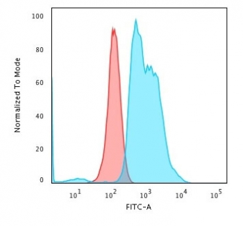
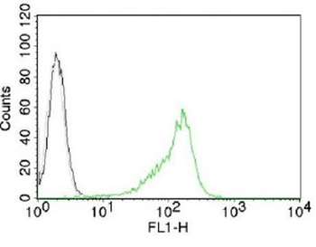
Intracellular FACS testing of 293 cells with Nucleoli marker antibody (green).
![Nucleoli Marker Antibody [NM95]](/images/pub/media/catalog/product/NewWebsite/15/orb1252651_5.jpg)
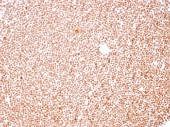
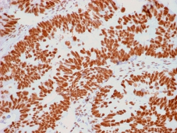
IHC staining of FFPE human colon carcinoma with Nucleoli Marker antibody. HIER: boil tissue sections in pH6, 10mM citrate buffer, for 10-20 min and allow to cool before testing.
Documents Download
Datasheet
Product Information
Request a Document
Protocol Information
IHC
Immunohistochemistry
FC
Flow Cytometry
IF
Immunofluorescence
ICC
Immunocytochemistry
Nucleoli Marker Antibody [NM95] (orb1252651)

- 0.0
Based on 0 reviews
Participating in our Biorbyt product reviews program enables you to support fellow scientists by sharing your firsthand experience with our products.
Login to Submit a ReviewAvailable Sizes
Select a size below
Choose Conjugation or Carrier Free Version
Free Secondary Antibody (20 ul)0/0
Please add an antibody product to your cart first.