You have no items in your shopping cart.
Featured
Description
Research Area
Musculoskeletal & Connective Tissue Research
Images & Validation
−Item 1 of 3
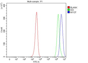
| Tested Applications | IF, IHC, WB |
|---|---|
| Reactivity | Human, Mouse, Rat |
| Application Notes |
Key Properties
−| Antibody Type | Primary Antibody |
|---|---|
| Host | Rabbit |
| Clonality | Polyclonal |
| Isotype | IgG |
| Immunogen | Recombinant fusion protein containing a sequence corresponding to amino acids 259-314 of human MYOT (NP_001129412.1). |
| Target | MYOT |
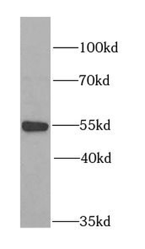
| Molecular Weight | Observed: 55kDa |
| Purification | Affinity purification |
| Conjugation | Unconjugated |
Storage & Handling
−| Storage | Maintain refrigerated at 2-8°C for up to 2 weeks. For long term storage store at -20°C in small aliquots to prevent freeze-thaw cycles. |
|---|---|
| Form/Appearance | Liquid |
| Buffer/Preservatives | PBS with 0.02% sodium azide, 50% glycerol, pH 7.3. |
| Concentration | batch dependent |
| Expiration Date | 12 months from date of receipt. |
| Disclaimer | For research use only |
Alternative Names
−MYOT, myotilin, LGMD1, LGMD1A, TTID, 57 kDa cytoskeletal protein, OTTHUMP00000159430, OTTHUMP00000223460, OTTHUMP00000223461, myofibrillar titin-like Ig domains protein, titin immunoglobulin domain protein (myotilin)
Similar Products
−- Item 1 of 4
MYOT Rabbit Polyclonal Antibody [orb1993151]
ELISA, FC, ICC, IHC, WB
Human, Mouse, Rat
Rabbit
Polyclonal
Unconjugated
100 μg - Item 1 of 2
- Item 1 of 2
- Item 1 of 2
MYOT Rabbit Polyclonal Antibody [orb629119]
ELISA, IF, IHC, WB
Human, Mouse, Rat
Rabbit
Polyclonal
Unconjugated
50 μg, 100 μg - Item 1 of 1


Quality Guarantee
Explore bioreagents carefree to elevate your research. All our products are rigorously tested for performance. If a product does not perform as described on its datasheet, our scientific support team will provide expert troubleshooting, a prompt replacement, or a refund. For full details, please see our Terms & Conditions and Buying Guide. Contact us at [email protected].
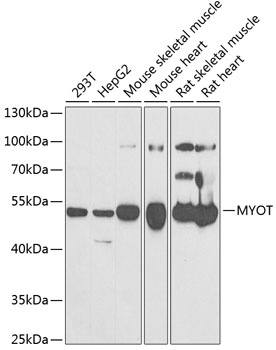
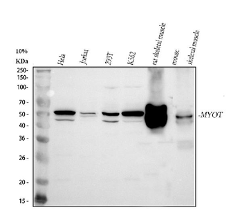
Western blot analysis of extracts of various cell lines, using MYOT antibody (orb1257010) at 1:1000 dilution. Secondary antibody: HRP Goat Anti-Rabbit IgG (H+L) at 1:10000 dilution. Lysates/proteins: 25 ug per lane. Blocking buffer: 3% nonfat dry milk in TBST. Detection: ECL Enhanced Kit. Exposure time: 90s.
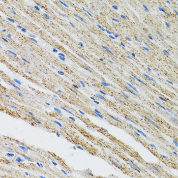
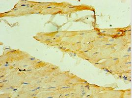
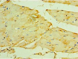
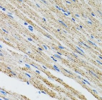
Immunohistochemistry of paraffin-embedded mouse heart using MYOT antibody (orb1257010) at dilution of 1:100 (40x lens).
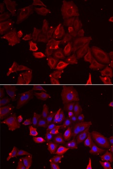
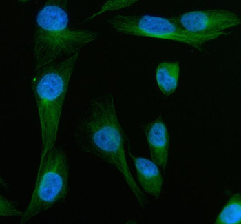
Immunofluorescence analysis of U2OS cells using MYOT antibody (orb1257010). Blue: DAPI for nuclear staining.
Quick Database Links
Gene Symbol
MYOT
UniProt
UniProt Details
− No UniProt data available
Documents Download
Datasheet
Product Information
Request a Document
Protocol Information
WB
Western Blot (IB, immunoblot)
IHC
Immunohistochemistry
IF
Immunofluorescence
MYOT Antibody (orb1257010)

- 0.0
Based on 0 reviews
Participating in our Biorbyt product reviews program enables you to support fellow scientists by sharing your firsthand experience with our products.
Login to Submit a ReviewAvailable Sizes
Select a size below
Choose Conjugation or Carrier Free Version
Free Secondary Antibody (20 ul)0/0
Please add an antibody product to your cart first.