You have no items in your shopping cart.
Description
Research Area
Signal Transduction
Images & Validation
−Item 1 of 5
| Tested Applications | FC, IHC-P, WB |
|---|---|
| Dilution Range | WB - 1:1000, IHC-P - 1:10-50, FC - 1:10-50 |
| Reactivity | Human |
| Predicted Reactivity | Mouse, Rat |
Key Properties
−| Host | Rabbit |
|---|---|
| Clonality | Polyclonal |
| Isotype | Rabbit IgG |
| Immunogen | This MUSK antibody is generated from rabbits immunized with a KLH conjugated synthetic peptide between 35-65 amino acids from the N-terminal region of human MUSK. Antigen Region: 35-65 aa. |
| Target | MUSK |
| Molecular Weight | 97056 Da |
| Conjugation | Unconjugated |
Storage & Handling
−| Storage | Maintain refrigerated at 2-8°C for up to 2 weeks. For long term storage store at -20°C in small aliquots to prevent freeze-thaw cycles |
|---|---|
| Form/Appearance | Purified polyclonal antibody supplied in PBS with 0.09% (W/V) sodium azide. This antibody is prepared by Saturated Ammonium Sulfate (SAS) precipitation followed by dialysis against PBS. |
| Expiration Date | 12 months from date of receipt. |
| Disclaimer | For research use only |
Alternative Names
−Muscle, skeletal receptor tyrosine-protein kinase, Muscle-specific tyrosine-protein kinase receptor, MuSK, Muscle-specific kinase receptor, MUSK
Quality Guarantee
Explore bioreagents carefree to elevate your research. All our products are rigorously tested for performance. If a product does not perform as described on its datasheet, our scientific support team will provide expert troubleshooting, a prompt replacement, or a refund. For full details, please see our Terms & Conditions and Buying Guide. Contact us at [email protected].
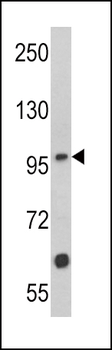
Western blot analysis of MUSK Antibody (N-term) in Jurkat cell line lysates (35 ug/lane). MUSK (arrow) was detected using the purified Pab.
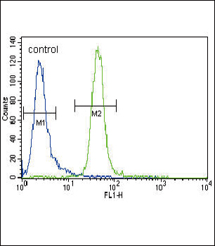
MUSK Antibody (N-term) flow cytometric analysis of CEM cells (right histogram) compared to a negative control cell (left histogram). FITC-conjugated goat-anti-rabbit secondary antibodies were used for the analysis.
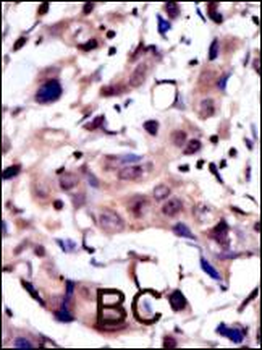
Formalin-fixed and paraffin-embedded human cancer tissue reacted with the primary antibody, which was peroxidase-conjugated to the secondary antibody, followed by AEC staining. This data demonstrates the use of this antibody for immunohistochemistry; clinical relevance has not been evaluated. BC = breast carcinoma; HC = hepatocarcinoma.
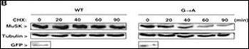
HA-tagged MuSK and GFP constructs were transfected into C2C12 cells. Transfected C2C12 cells were treated with cycloheximide (CHX) and at different times after addition of CHX, amounts of WT and mutant MuSK were analyzed by WB with anti-HA antibody. Alpha-tubulin was used as an internal control and transfection efficiency was verified with anti-GFP antibody.
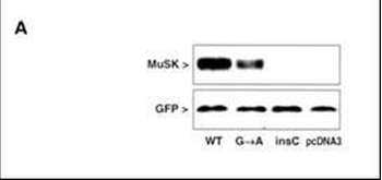
MuSK protein expression in extracts of COS cells after transfection with MuSK mutated and GFP constructs. WB with polyclonal MuSK and monoclonal GFP antibodies showed normal expression of the wild-type MuSK protein (WT), diminished expression of the GA mutant MuSK and no expression of the insC mutant or the pcDNA3 vector alone in transfected COS cells. GFP cotransfection was used to verify transfection efficiency.
Quick Database Links
UniProt Details
− No UniProt data available
NCBI Reference Sequences
−Associated Accession Numbers
Curated reference sequences for the gene transcript and protein product| Protein | NP_005583.1, NP_001159752.1, NP_001159753.1 |
|---|
Documents Download
Datasheet
Product Information
Request a Document
Protocol Information
WB
Western Blot (IB, immunoblot)
IHC-P
Immunohistochemistry Paraffin
FC
Flow Cytometry
MUSK Antibody (N-term) (orb1929012)

- 0.0
Based on 0 reviews
Participating in our Biorbyt product reviews program enables you to support fellow scientists by sharing your firsthand experience with our products.
Login to Submit a ReviewAvailable Sizes
Select a size below
Free Secondary Antibody (20 ul)0/0
Please add an antibody product to your cart first.