You have no items in your shopping cart.
Description
Research Area
Epigenetics & Chromatin
Images & Validation
−Item 1 of 8
| Tested Applications | FACS, IF, IHC-P, WB |
|---|---|
| Dilution Range | Flow cytometry: 1-2ug/million cells in 0.1ml,Western blot: 1-2ug/ml,Immunohistochemistry (FFPE): 1-2ug/ml,Immunofluorescence: 1-2ug/ml |
| Reactivity | Human |
| Application Notes |
Key Properties
−| Antibody Type | Primary Antibody |
|---|---|
| Host | Mouse |
| Clonality | Monoclonal |
| Isotype | Mouse IgG1, kappa |
| Clone No. | MSH2/2622 |
| Immunogen | A recombinant human partial protein (amino acids 327-427) was used as the immunogen for this MSH2 antibody. |
| Purification | Protein G affinity chromatography |
| Conjugation | Unconjugated |
Storage & Handling
−| Storage | Maintain refrigerated at 2-8°C for up to 2 weeks. For long term storage store at -20°C in small aliquots to prevent freeze-thaw cycles. |
|---|---|
| Buffer/Preservatives | 0.2 mg/ml in 1X PBS with 0.1 mg/ml rAlbumin and 0.05% sodium azide |
| Expiration Date | 12 months from date of receipt. |
| Disclaimer | For research use only |
Similar Products
−- Item 1 of 9
- Item 1 of 8
- Item 1 of 8
- Item 1 of 6
MSH2 Rabbit Polyclonal Antibody [orb234330]
FC, ICC, IF, IHC, WB
Human, Mouse, Rat
Rabbit
Polyclonal
Unconjugated
100 μg - Item 1 of 7


Quality Guarantee
Explore bioreagents carefree to elevate your research. All our products are rigorously tested for performance. If a product does not perform as described on its datasheet, our scientific support team will provide expert troubleshooting, a prompt replacement, or a refund. For full details, please see our Terms & Conditions and Buying Guide. Contact us at [email protected].
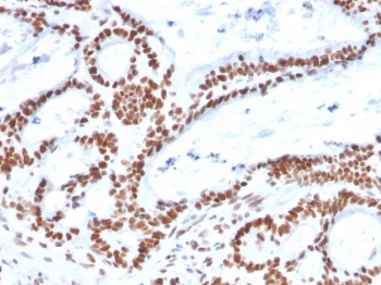
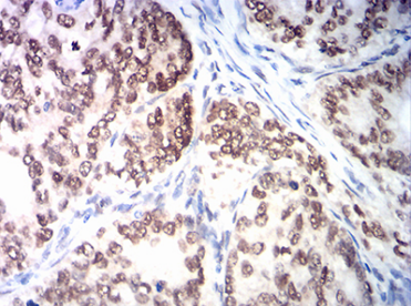
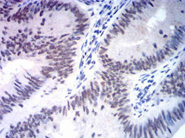
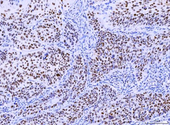
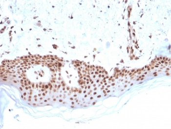
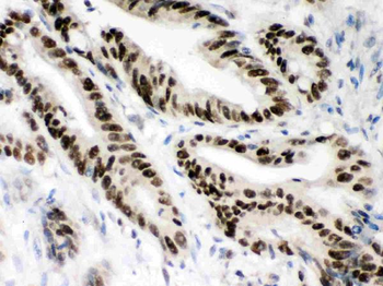
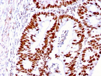
IHC staining of FFPE human colon carcinoma with MSH2 antibody. HIER: boil tissue sections in pH9 10mM Tris with 1mM EDTA for 20 min and allow to cool before testing.

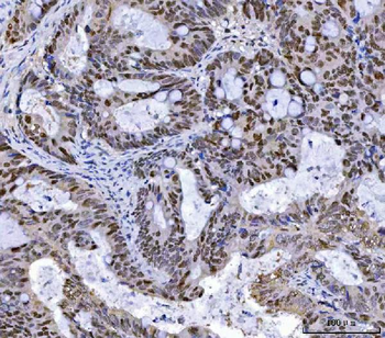
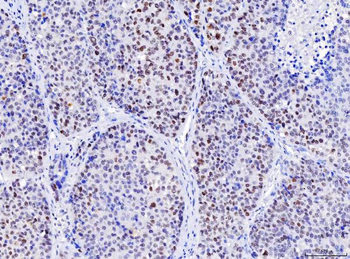
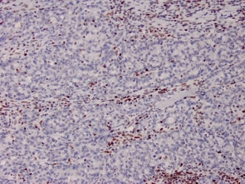
IHC staining of FFPE human basal cell carcinoma with MSH2 antibody. HIER: boil tissue sections in pH9 10mM Tris with 1mM EDTA for 20 min and allow to cool before testing.
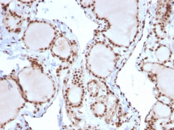
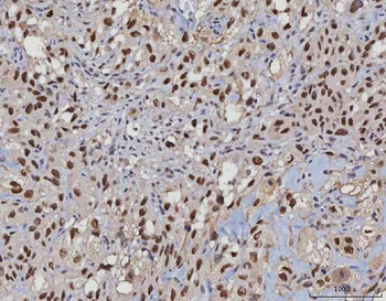
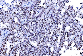
IHC staining of FFPE human thyroid carcinoma with MSH2 antibody. HIER: boil tissue sections in pH9 10mM Tris with 1mM EDTA for 20 min and allow to cool before testing.
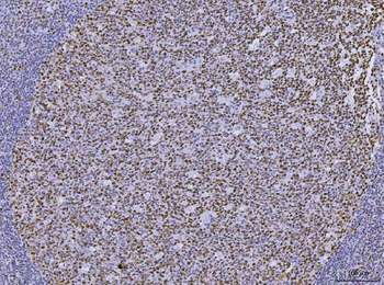
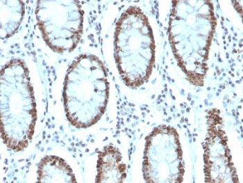
IHC staining of FFPE human colon from a Lynch disease patient with MSH2 antibody. HIER: boil tissue sections in pH9 10mM Tris with 1mM EDTA for 20 min and allow to cool before testing.
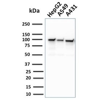
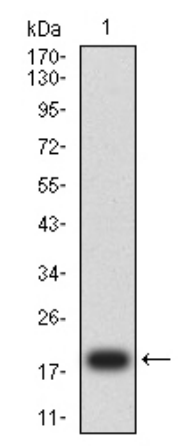
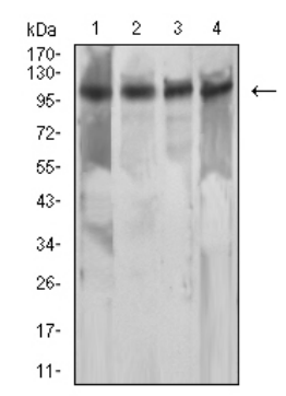
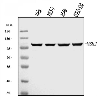
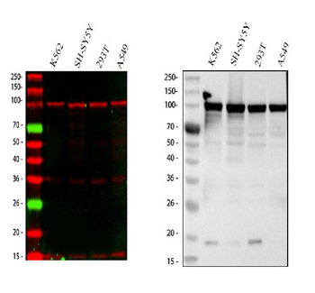
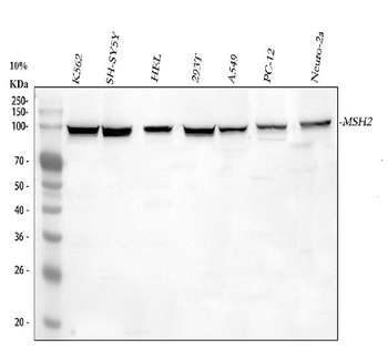
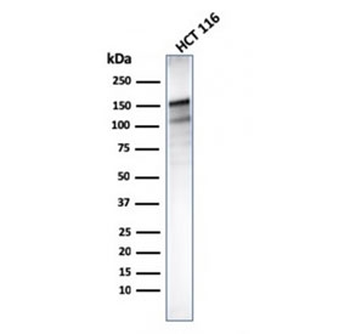
Western blot testing of human cell lysates with MSH2 antibody. Expected molecular weight: ~105 kDa.
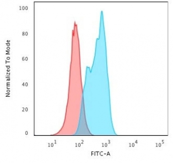
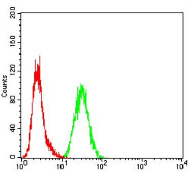
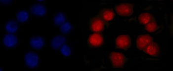
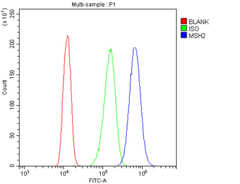
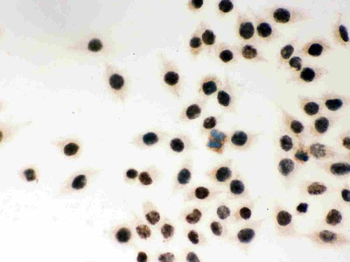
Flow cytometry testing of permeabilized human A549 cells with MSH2 antibody; Red=isotype control, Blue= MSH2 antibody.

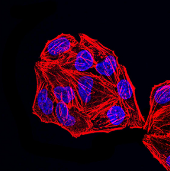
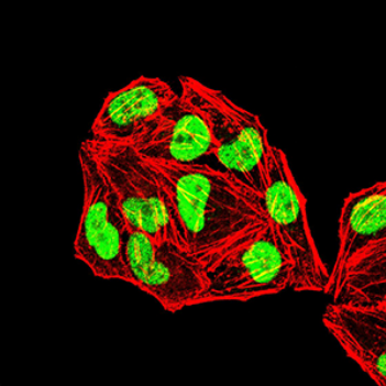
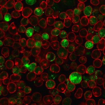
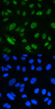
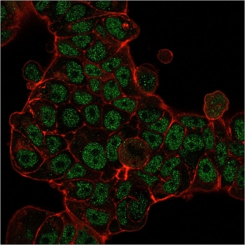
Immunofluorescent staining of permeabilized human MOLT-4 cells with MSH2 antibody (green) and Phalloidin (red).
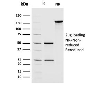
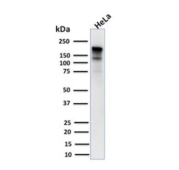
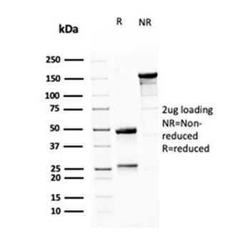
SDS-PAGE analysis of purified, BSA-free MSH2 antibody as confirmation of integrity and purity.
Quick Database Links
UniProt
UniProt Details
− No UniProt data available
Documents Download
Datasheet
Product Information
Request a Document
Protocol Information
WB
Western Blot (IB, immunoblot)
IHC-P
Immunohistochemistry Paraffin
FACS
Fluorescence-Activated Cell Sorting (FC, Flow cytometry)
IF
Immunofluorescence
MSH2 Antibody (orb639818)

- 0.0
Based on 0 reviews
Participating in our Biorbyt product reviews program enables you to support fellow scientists by sharing your firsthand experience with our products.
Login to Submit a ReviewAvailable Sizes
Select a size below
Choose Conjugation or Carrier Free Version
Free Secondary Antibody (20 ul)0/0
Please add an antibody product to your cart first.