You have no items in your shopping cart.
Description
Research Area
Cancer Biology
Images & Validation
−Item 1 of 3
| Tested Applications | IP, WB |
|---|---|
| Dilution Range | WB - 1:1000; IP - 20 µl/mg lysate |
| Reactivity | Human, Mouse |
| Application Notes |
Key Properties
−| Antibody Type | Primary Antibody |
|---|---|
| Host | Mouse |
| Clonality | Monoclonal |
| Isotype | IgG1, kappa |
| Clone No. | 2A1-9A4 |
| Immunogen | Amino terminal one-third of the CIP2A (GeneID 57650) recombinant protein |
| Target | CIP2A |
| Purification | Purified |
| Conjugation | Unconjugated |
Storage & Handling
−| Storage | 2 - 8°C |
|---|---|
| Form/Appearance | Liquid |
| Buffer/Preservatives | Tris-buffered Saline containing 0.1% rAlbumin and 0.09% Sodium Azide |
| Concentration | 40 µg/ml |
| Expiration Date | 12 months from date of receipt. |
| Disclaimer | For research use only |
Alternative Names
−cancerous inhibitor of PP2A; cancerous inhibitor of protein phosphatase 2A; KIAA1524; p90; p90 autoantigen; protein CIP2A
Similar Products
−- Item 1 of 1
CIP2A Mouse Monoclonal Antibody [orb1292535]
ELISA, WB
Human, Rat
Mouse
Monoclonal
Unconjugated
50 μg, 100 μg 
Mouse CIP2A Monoclonal Antibody [orb1520148]
IP, WB
Human, Mouse
Mouse
Monoclonal
Unconjugated
100 μg (BSA-free)
Quality Guarantee
Explore bioreagents carefree to elevate your research. All our products are rigorously tested for performance. If a product does not perform as described on its datasheet, our scientific support team will provide expert troubleshooting, a prompt replacement, or a refund. For full details, please see our Terms & Conditions and Buying Guide. Contact us at [email protected].

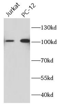
Detection of human CIP2A by western blot. Samples: Whole cell lysate (25 µg) from HEK293T, Jurkat, U2OS, HeLa, and A-549 cells prepared using NETN lysis buffer. Antibody: Mouse anti-CIP2A monoclonal antibody [2A1-9A4] (orb1520149) used at 1:1000. Secondary: HRP-conjugated goat anti-mouse IgG. Detection: Chemiluminescence with an exposure time of 75 seconds. Lower Panel: Rabbit anti-COPB2 antibody.
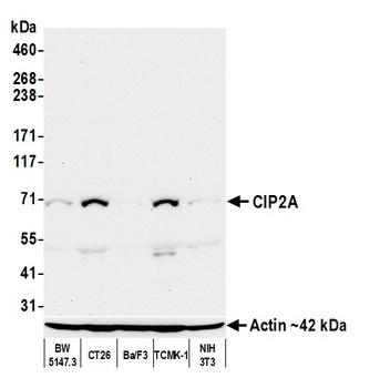
Detection of mouse CIP2A by western blot. Samples: Whole cell lysate (50 µg) from BW5147.3, CT26, Ba/F3, TCMK-1, and NIH 3T3 cells prepared using NETN lysis buffer. Antibody: Mouse anti-CIP2A monoclonal antibody [2A1-9A4] (orb1520149) used at 1:1000. Secondary: HRP-conjugated goat anti-mouse IgG. Detection: Chemiluminescence with an exposure time of 75 seconds. Lower Panel: Rabbit anti-Actin recombinant monoclonal antibody.
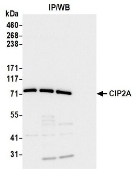
Detection of human CIP2A by western blot of immunoprecipitates. Samples: Whole cell lysate (1.0 mg per IP reaction; 20% of IP loaded) from HEK293T cells prepared using NETN lysis buffer. Antibodies: Mouse anti-CIP2A monoclonal antibody [2A1-9A4] (orb1520149) used for IP at 20 µl/mg lysate.
Quick Database Links
UniProt Details
− No UniProt data available
NCBI Reference Sequences
−Associated Accession Numbers
Curated reference sequences for the gene transcript and protein product| Protein | NP_065941.1 |
|---|
Documents Download
Datasheet
Product Information
Request a Document
Protocol Information
WB
Western Blot (IB, immunoblot)
IP
Immunoprecipitation
Mouse CIP2A Monoclonal Antibody (orb1520149)

- 0.0
Based on 0 reviews
Participating in our Biorbyt product reviews program enables you to support fellow scientists by sharing your firsthand experience with our products.
Login to Submit a ReviewAvailable Sizes
Select a size below
Choose Conjugation or Carrier Free Version
Free Secondary Antibody (20 ul)0/0
Please add an antibody product to your cart first.