You have no items in your shopping cart.
Featured
Description
Research Area
Signal Transduction
Images & Validation
−Item 1 of 3
| Tested Applications | ICC, IF, IHC, IP, WB |
|---|---|
| Dilution Range | WB (1:1000), IHC (1:1000), ICC/IF (1:100) |
| Reactivity | Human, Mouse, Rat |
| Application Notes |
Key Properties
−| Host | Mouse |
|---|---|
| Clonality | Monoclonal |
| Isotype | IgG1 |
| Clone No. | N75/33 (Formerly sold as S75-33) |
| Immunogen | Fusion protein amino acids 824-1203 (cytoplasmic C-terminus) of rat mGluR5b |
| Target | mGluR5 |
| Molecular Weight | 130kDa |
| Purification | Protein G Purified |
| Conjugation | APC |
Storage & Handling
−| Storage | Conjugated antibodies should be stored according to the product label |
|---|---|
| Buffer/Preservatives | 95.46mM Phosphate, 2.48mM MES and 2mM EDTA |
| Concentration | 1 mg/ml |
| Expiration Date | 12 months from date of receipt. |
| Disclaimer | For research use only |
Alternative Names
−mGluR5, mGlu5, GRM5, GPRC1E, Metabotropic glutamate receptor 5, Metabotropic glutamate receptor 5 variant F, Metabotropic glutamate receptor 5 variant G, Metabotropic glutamate receptor 5 variant H, Glutamate receptor metabotropic 5, mGluR5a, mGluR5b, mGluR, Glutamate receptor metabotropic 1, mGlu1, mGluR1, GRM1, GRM 1, GRM1A, GRM1 alpha, GRM1_HUMAN, GPRC1A, Metabotropic glutamate receptor 1, mGluR1 alpha
Similar Products
−- Item 1 of 3

MGluR5 Rabbit Polyclonal Antibody (APC-Cy5.5) [orb2534111]
IF
Canine, Gallus, Human, Mouse, Sheep
Rat
Rabbit
Polyclonal
APC/Cy5.5
100 μlMGluR5 Rabbit Polyclonal Antibody (APC-Cy7) [orb2534110]
IF
Canine, Gallus, Human, Mouse, Sheep
Rat
Rabbit
Polyclonal
APC/Cy7
100 μlMGluR5 Rabbit Polyclonal Antibody (APC) [orb995464]
IF
Canine, Gallus, Human, Mouse, Sheep
Rat
Rabbit
Polyclonal
APC
100 μlMetabotropic Glutamate Receptor 5/GRM5 Rabbit Polyclonal Antibody (APC) [orb2608544]
FC
Human, Mouse, Rat
Rabbit
Polyclonal
APC
100 μg
Quality Guarantee
Explore bioreagents carefree to elevate your research. All our products are rigorously tested for performance. If a product does not perform as described on its datasheet, our scientific support team will provide expert troubleshooting, a prompt replacement, or a refund. For full details, please see our Terms & Conditions and Buying Guide. Contact us at [email protected].
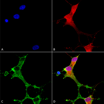
Immunocytochemistry/Immunofluorescence analysis using Mouse Anti-mGluR1/5 Monoclonal Antibody, Clone N75/33. Tissue: Neuroblastoma cells (SH-SY5Y). Species: Human. Fixation: 4% PFA for 15 min. Primary Antibody: Mouse Anti-mGluR1/5 Monoclonal Antibody at 1:100 for overnight at 4°C with slow rocking. Secondary Antibody: AlexaFluor 488 at 1:1000 for 1 hour at RT. Counterstain: Phalloidin-iFluor 647 (red) F-Actin stain; Hoechst (blue) nuclear stain at 1:800, 1.6mM for 20 min at RT. (A) Hoechst (blue) nuclear stain. (B) Phalloidin-iFluor 647 (red) F-Actin stain. (C) mGluR1/5 Antibody (D) Composite.
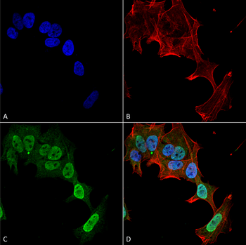
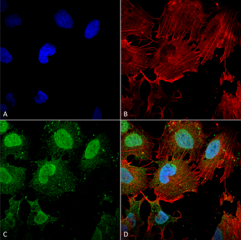
Immunocytochemistry/Immunofluorescence analysis using Mouse Anti-mGluR1/5 Monoclonal Antibody, Clone N75/33. Tissue: Neuroblastoma cell line (SK-N-BE). Species: Human. Fixation: 4% Formaldehyde for 15 min at RT. Primary Antibody: Mouse Anti-mGluR1/5 Monoclonal Antibody at 1:100 for 60 min at RT. Secondary Antibody: Goat Anti-Mouse ATTO 488 at 1:200 for 60 min at RT. Counterstain: Phalloidin Texas Red F-Actin stain; DAPI (blue) nuclear stain at 1:1000, 1:5000 for 60 min at RT, 5 min at RT. Localization: Cell Membrane, Cytoplasm, Nucleus. Magnification: 60X. (A) DAPI (blue) nuclear stain. (B) Phalloidin Texas Red F-Actin stain. (C) mGluR1/5 Antibody. (D) Composite.
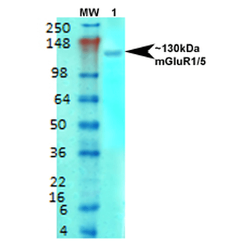
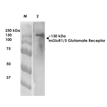
Western Blot analysis of Rat brain membrane lysate showing detection of mGluR5 Glutamate Receptor protein using Mouse Anti-mGluR5 Glutamate Receptor Monoclonal Antibody, Clone N75/33. Primary Antibody: Mouse Anti-mGluR5 Glutamate Receptor Monoclonal Antibody at 1:1000.
Quick Database Links
UniProt Details
− No UniProt data available
NCBI Gene Details
− No NCBI Gene data available
NCBI Reference Sequences
−Associated Accession Numbers
Curated reference sequences for the gene transcript and protein product| Protein | NP_058708.1 |
|---|
Documents Download
Datasheet
Product Information
Request a Document
Protocol Information
WB
Western Blot (IB, immunoblot)
IHC
Immunohistochemistry
IF
Immunofluorescence
ICC
Immunocytochemistry
IP
Immunoprecipitation
mGluR5 Antibody (APC) (orb149945)

- 0.0
Based on 0 reviews
Participating in our Biorbyt product reviews program enables you to support fellow scientists by sharing your firsthand experience with our products.
Login to Submit a ReviewAvailable Sizes
Select a size below
Choose Conjugation or Carrier Free Version
Free Secondary Antibody (20 ul)0/0
Please add an antibody product to your cart first.