You have no items in your shopping cart.
Description
Research Area
Cancer Biology
Images & Validation
−Item 1 of 2
| Tested Applications | IHC-P, WB |
|---|---|
| Dilution Range | Western blot: 1-2ug/ml,Immunohistochemistry (FFPE): 1-2ug/ml for 30 min at RT |
| Reactivity | Human |
| Application Notes |
Key Properties
−| Antibody Type | Primary Antibody |
|---|---|
| Host | Mouse |
| Clonality | Monoclonal |
| Isotype | Mouse IgG1 + IgG2a + IgG2b + IgG2b |
| Clone No. | M2-7C10 + M2-9E3 + HMB45 + T311 |
| Immunogen | Recombinant MART-1 protein (M2-7C10 & M2-9E3), recombinant tyrosinase protein (T311) and extract of pigmented melanoma metastases from lymph nodes (HMB45) were used as the immunogens for this Melanoma Cell Marker antibody cocktail. |
| Purification | Protein G affinity chromatography |
| Conjugation | Unconjugated |
Storage & Handling
−| Storage | Maintain refrigerated at 2-8°C for up to 2 weeks. For long term storage store at -20°C in small aliquots to prevent freeze-thaw cycles. |
|---|---|
| Buffer/Preservatives | 0.2 mg/ml in 1X PBS with 0.1 mg/ml rAlbumin and 0.05% sodium azide |
| Expiration Date | 12 months from date of receipt. |
| Disclaimer | For research use only |
Similar Products
−- Item 1 of 2
- Item 1 of 2
Melanoma Marker Antibody Cocktail (MART-1 & gp100) [orb640117]
IHC-P
Human
Mouse
Monoclonal
Unconjugated
20 μg, 100 μg - Item 1 of 2
MART-1 Antibody Cocktail [orb248351]
FACS, IF, IHC-P, WB
Human, Mouse, Rat
Mouse
Monoclonal
Unconjugated
100 μg, 20 μg - Item 1 of 2
- Item 1 of 2
Quality Guarantee
Explore bioreagents carefree to elevate your research. All our products are rigorously tested for performance. If a product does not perform as described on its datasheet, our scientific support team will provide expert troubleshooting, a prompt replacement, or a refund. For full details, please see our Terms & Conditions and Buying Guide. Contact us at [email protected].
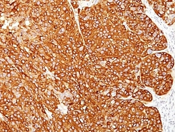
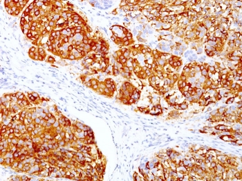
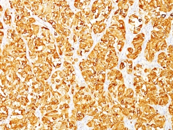
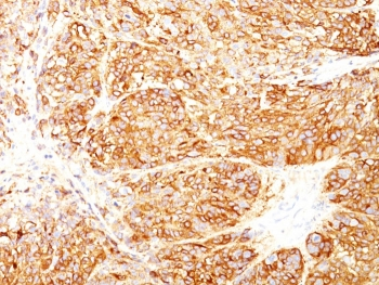
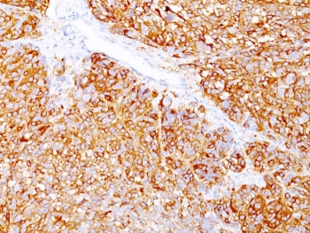
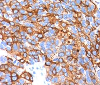
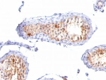
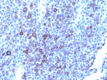
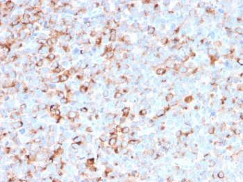
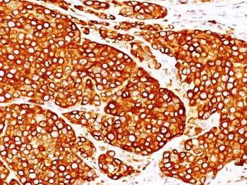
IHC: Formalin-fixed, paraffin-embedded human melanoma stained with melanoma Marker antibody cocktail (M2-7C10 + M2-9E3 + T311 + HMB45).
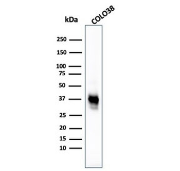
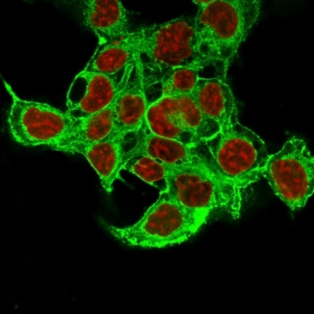
Western blot testing of human COLO-38 cell lysate with Melanoma Cell Marker antibody cocktail (clones M2-7C10 + M2-9E3 + HMB45 + T311).
Documents Download
Datasheet
Product Information
Request a Document
Protocol Information
WB
Western Blot (IB, immunoblot)
IHC-P
Immunohistochemistry Paraffin
Melanoma Cell Marker Antibody Cocktail (orb385745)

- 0.0
Based on 0 reviews
Participating in our Biorbyt product reviews program enables you to support fellow scientists by sharing your firsthand experience with our products.
Login to Submit a ReviewAvailable Sizes
Select a size below
Choose Conjugation or Carrier Free Version
Free Secondary Antibody (20 ul)0/0
Please add an antibody product to your cart first.