You have no items in your shopping cart.
Featured
Description
Research Area
Epigenetics & Chromatin
Images & Validation
−Item 1 of 3
| Tested Applications | ICC, IF, WB |
|---|---|
| Dilution Range | WB (1:2000), ICC/IF (1:100) |
| Reactivity | Bovine, Human, Mouse, Primate |
| Application Notes |
Key Properties
−| Host | Mouse |
|---|---|
| Clonality | Monoclonal |
| Isotype | IgG1 |
| Clone No. | P2B11 |
| Immunogen | GST-tagged recombinant protein corresponding to mouse MDC1 at and around the N-terminus |
| Target | MDC1 |
| Molecular Weight | 184kDa |
| Purification | Protein G Purified |
| Conjugation | Unconjugated |
Storage & Handling
−| Storage | Maintain refrigerated at 2-8°C for up to 2 weeks. For long term storage store at -20°C in small aliquots to prevent freeze-thaw cycles. |
|---|---|
| Buffer/Preservatives | PBS pH 7.4, 50% glycerol, 0.09% sodium azide. Storage buffer changes when conjugated. |
| Concentration | 1 mg/ml |
| Expiration Date | 12 months from date of receipt. |
| Disclaimer | For research use only |
Alternative Names
−MDC1, Nuclear factor with BRCT domains1, Mediator of DNA damage checkpoint 1
Similar Products
−- Item 1 of 7
- Item 1 of 5
MDC1 Rabbit Polyclonal Antibody [orb402240]
ELISA, FC, ICC, IF, IHC
Human
Rabbit
Polyclonal
Unconjugated
100 μg - Item 1 of 2
- Item 1 of 2
MDC1 Rabbit Polyclonal Antibody [orb628707]
ELISA, IHC, WB
Human
Rabbit
Polyclonal
Unconjugated
50 μg, 100 μg - Item 1 of 2


Quality Guarantee
Explore bioreagents carefree to elevate your research. All our products are rigorously tested for performance. If a product does not perform as described on its datasheet, our scientific support team will provide expert troubleshooting, a prompt replacement, or a refund. For full details, please see our Terms & Conditions and Buying Guide. Contact us at [email protected].
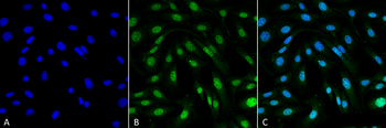
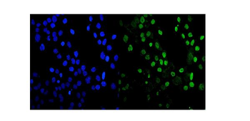
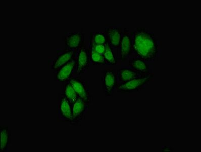
Immunocytochemistry/Immunofluorescence analysis using Mouse Anti-MDC1 Monoclonal Antibody, Clone P2B11. Tissue: Fibroblast cell line (NIH 3T3). Species: Mouse. Fixation: 4% Formaldehyde for 15 min at RT. Primary Antibody: Mouse Anti-MDC1 Monoclonal Antibody at 1:100 for 60 min at RT. Secondary Antibody: Goat Anti-Mouse ATTO 488 at 1:100 for 60 min at RT. Counterstain: DAPI (blue) nuclear stain at 1:5000 for 5 min RT. Localization: Nucleus. Magnification: 60X.
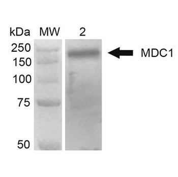
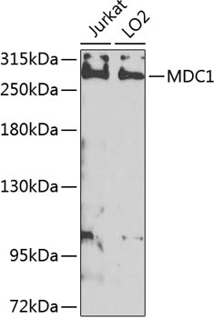
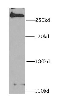
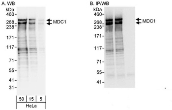
Western Blot analysis of Human Embryonic kidney epithelial cell line (HEK293T) lysate showing detection of 184 kDa MDC1 protein using Mouse Anti-MDC1 Monoclonal Antibody, Clone P2B11. Lane 1: MW ladder. Lane 2: 293Trap cell lysates. Load: 30 μg. Block: 5% Skim Milk in 1X TBST. Primary Antibody: Mouse Anti-MDC1 Monoclonal Antibody at 1:1000 for 2 hours RT. Secondary Antibody: Goat Anti-Mouse HRP: IgG at 1:2000 for 60 min at RT. Color Development: ECL solution for 5 min in RT. Predicted/Observed Size: 184 kDa.
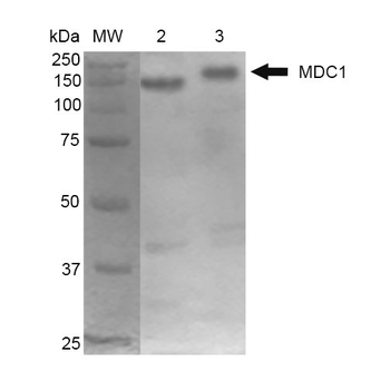
Western Blot analysis of Mouse Cortex and Cerebellum showing detection of 184 kDa MDC1 protein using Mouse Anti-MDC1 Monoclonal Antibody, Clone P2B11. Lane 1: MW ladder. Lane 2: Mouse Cortex. Lane 3: Mouse Cerebellum. Load: 10 μg. Block: 5% Skim Milk in 1X TBST. Primary Antibody: Mouse Anti-MDC1 Monoclonal Antibody at 1:1000 for 2 hours RT. Secondary Antibody: Goat Anti-Mouse at 1:2000 for 60 min at RT. Color Development: ECL solution for 5 min in RT. Predicted/Observed Size: 184 kDa.
Quick Database Links
UniProt Details
− No UniProt data available
NCBI Gene Details
− No NCBI Gene data available
NCBI Reference Sequences
−Associated Accession Numbers
Curated reference sequences for the gene transcript and protein product| Protein | NP_001010833.2 |
|---|
Documents Download
Datasheet
Product Information
Request a Document
Protocol Information
WB
Western Blot (IB, immunoblot)
IF
Immunofluorescence
ICC
Immunocytochemistry
MDC1 Antibody (orb67385)

- 0.0
Based on 0 reviews
Participating in our Biorbyt product reviews program enables you to support fellow scientists by sharing your firsthand experience with our products.
Login to Submit a ReviewAvailable Sizes
Select a size below
Choose Conjugation or Carrier Free Version
Free Secondary Antibody (20 ul)0/0
Please add an antibody product to your cart first.