You have no items in your shopping cart.
MAP2 Antibody
SKU: orb1925242
Description
Images & Validation
−Item 1 of 4
| Tested Applications | FC, IHC-P, WB |
|---|---|
| Dilution Range | WB - 1:1000, IHC-P - 1:100-500, FC - 1:10-50 |
| Reactivity | Human, Rat |
Key Properties
−| Antibody Type | Primary Antibody |
|---|---|
| Host | Rabbit |
| Clonality | Polyclonal |
| Isotype | Rabbit IgG |
| Molecular Weight | 199526 Da |
| Conjugation | Unconjugated |
Storage & Handling
−| Storage | Maintain refrigerated at 2-8°C for up to 2 weeks. For long term storage store at -20°C in small aliquots to prevent freeze-thaw cycles |
|---|---|
| Form/Appearance | Purified polyclonal antibody supplied in PBS with 0.05% (V/V) Proclin 300. This antibody is prepared by Saturated Ammonium Sulfate (SAS) precipitation followed by dialysis against PBS. |
| Expiration Date | 12 months from date of receipt. |
| Disclaimer | For research use only |
Similar Products
−- Item 1 of 16
Phospho-ERK1/2 (Thr202 + Tyr204) Rabbit Polyclonal Antibody [orb5178]
FC, ICC, IF, IHC-Fr, IHC-P
Bovine, Canine, Equine, Gallus, Guinea pig, Porcine, Rabbit
Human, Mouse, Rat
Rabbit
Polyclonal
Unconjugated
50 μl, 100 μl, 200 μl - Item 1 of 10
Phospho-ERK1 (Thr202/Tyr204) + ERK2 (Thr183/Tyr185) Rabbit Polyclonal Antibody [orb783430]
FC, IF, IHC
Bovine, Canine, Equine, Gallus, Guinea pig, Porcine, Rabbit
Human, Mouse, Rat
Rabbit
Polyclonal
Unconjugated
50 μl, 100 μl, 200 μl - Item 1 of 7
MAP2 Rabbit Polyclonal Antibody [orb11455]
FC, IF, IHC-Fr, IHC-P, WB
Mouse, Rat
Human, Mouse, Rat
Rabbit
Polyclonal
Unconjugated
100 μl, 50 μl, 200 μl - Item 1 of 6
ERK1 + ERK2 Rabbit Polyclonal Antibody [orb10604]
FC, IF, IHC-Fr, IHC-P, WB
Bovine, Canine, Equine, Gallus, Goat, Porcine, Rabbit, Sheep
Human, Mouse, Rat
Rabbit
Polyclonal
Unconjugated
200 μl, 50 μl, 100 μl - Item 1 of 7
ERK1/2 Recombinant Rabbit Monoclonal Antibody [orb704524]
FC, ICC, IF, IHC-Fr, IHC-P, WB
Zebrafish
Human, Mouse, Rat
Rabbit
Recombinant
Unconjugated
50 μl, 100 μl, 25 μl


Quality Guarantee
Explore bioreagents carefree to elevate your research. All our products are rigorously tested for performance. If a product does not perform as described on its datasheet, our scientific support team will provide expert troubleshooting, a prompt replacement, or a refund. For full details, please see our Terms & Conditions and Buying Guide. Contact us at [email protected].
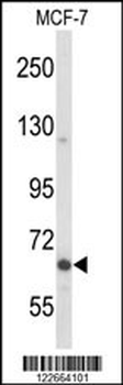
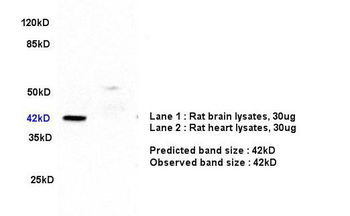
Western blot analysis of MAP2 Antibody in MCF-7 cell line lysates (35 ug/lane). MAP2 (arrow) was detected using the purified Pab.
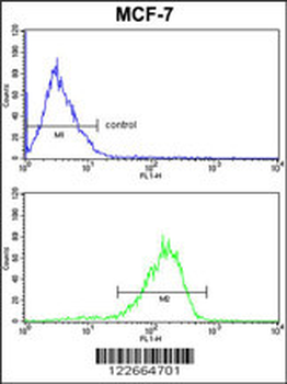
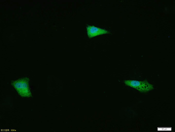
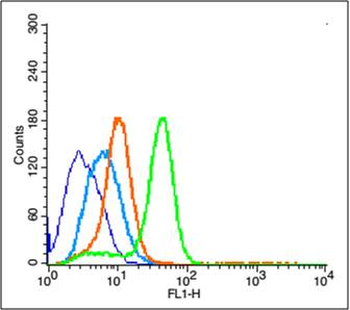
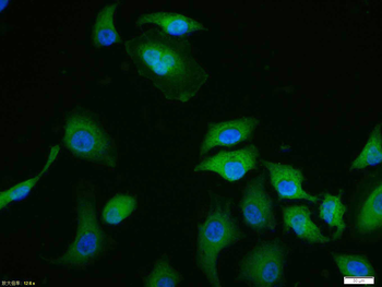
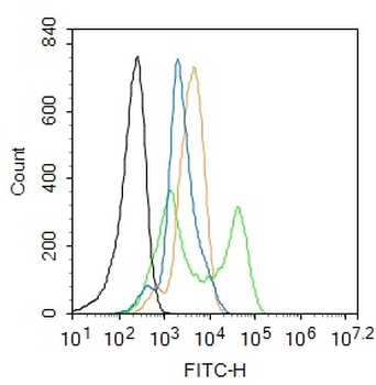
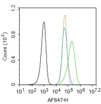
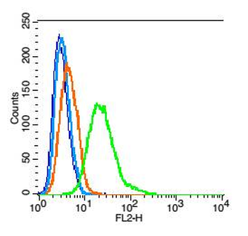
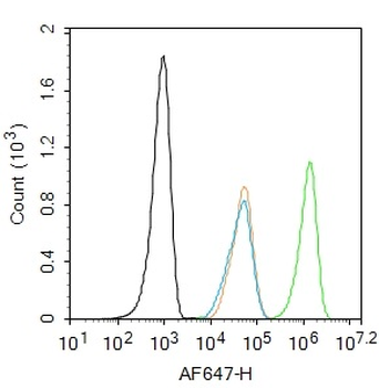
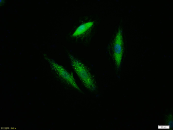
MAP2 Antibody FC analysis of MCF-7 cells (bottom histogram) compared to a negative control cell (top histogram). FITC-conjugated goat-anti-rabbit secondary antibodies were used for the analysis.
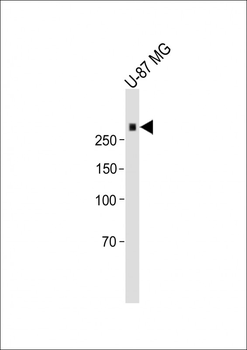
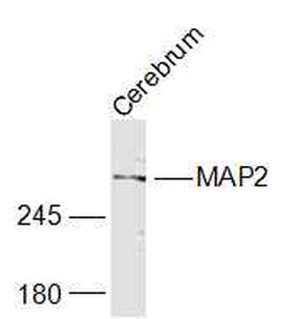
All lanes: Anti-MAP2 Antibody at 1:1000 dilution+ U-87 MG Cell lysate. Lysates/proteins at 20 µg per lane. Secondary Goat Anti-rabbit IgG, (H+L), Peroxidase conjugated at 1/15000 dilution.Observed band size: 280kDa. Blocking/Dilution buffer: 5% NFDM/TBST.
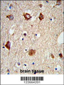
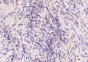
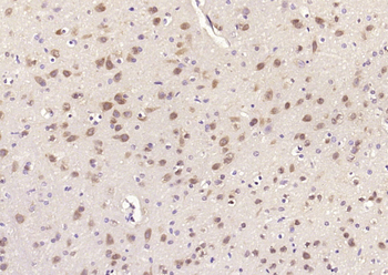
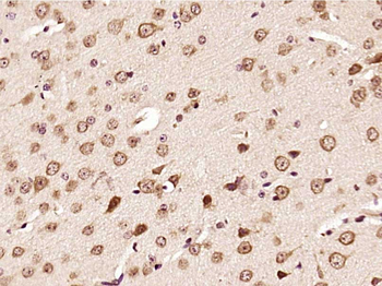
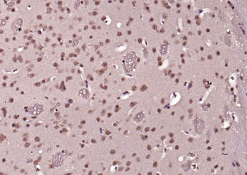
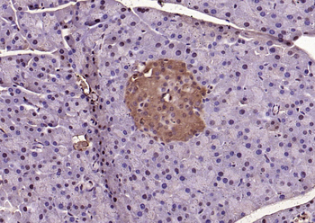
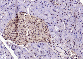
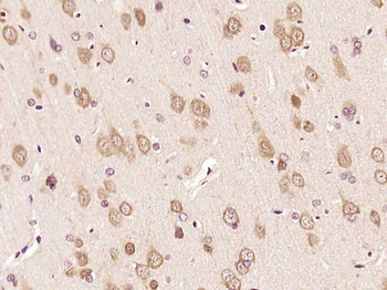
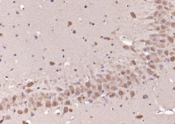
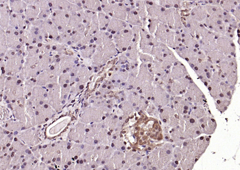
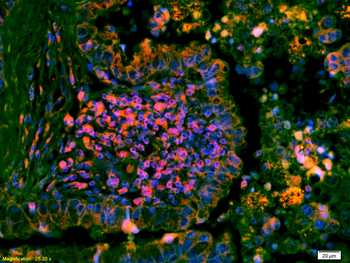
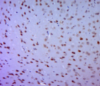
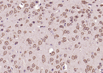
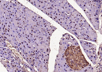
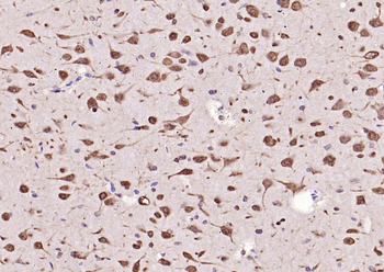
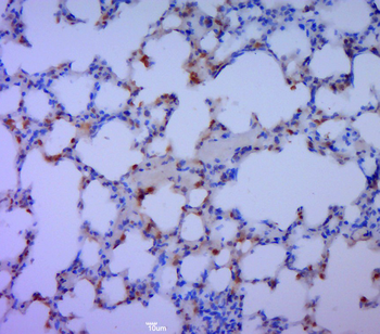
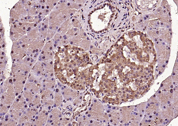
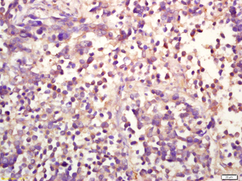
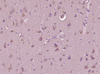
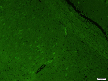
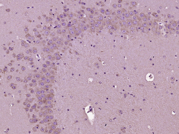
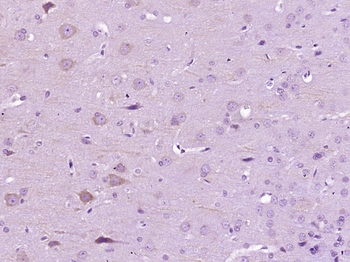
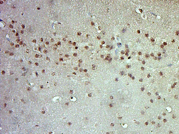
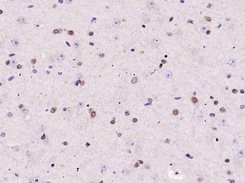
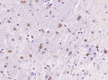
Formalin-fixed and paraffin-embedded mouse brain tissue reacted with MAP2 Antibody, which was peroxidase-conjugated to the secondary antibody, followed by DAB staining. This data demonstrates the use of this antibody for immunohistochemistry; clinical relevance has not been evaluated.
Quick Database Links
UniProt
UniProt Details
− No UniProt data available
Documents Download
Datasheet
Product Information
Request a Document
Protocol Information
WB
Western Blot (IB, immunoblot)
IHC-P
Immunohistochemistry Paraffin
FC
Flow Cytometry
MAP2 Antibody (orb1925242)

- 0.0
Based on 0 reviews
Participating in our Biorbyt product reviews program enables you to support fellow scientists by sharing your firsthand experience with our products.
Login to Submit a ReviewAvailable Sizes
Select a size below