You have no items in your shopping cart.
Featured
Description
Research Area
Signal Transduction
Images & Validation
−Item 1 of 2
| Tested Applications | ICC, IF, WB |
|---|---|
| Dilution Range | WB (1:1000) |
| Reactivity | Human, Mouse, Rat |
| Application Notes |
Key Properties
−| Host | Mouse |
|---|---|
| Clonality | Monoclonal |
| Isotype | IgG2a |
| Clone No. | N165/38 (Formerly sold as S165-38) |
| Immunogen | Fusion protein amino acids 1315-1607 (cytoplasmic C-terminus) of human LAR. 97% identical in both rat and mouse. > 80% identity with PTPRD and PTPRS. > 50% identity with PTPRM and PTPRK. |
| Target | LAR |
| Molecular Weight | 85kDa |
| Purification | Protein G Purified |
| Conjugation | PerCP |
Storage & Handling
−| Storage | Conjugated antibodies should be stored according to the product label |
|---|---|
| Buffer/Preservatives | 95.46mM Phosphate, 2.48mM MES and 2mM EDTA |
| Concentration | 1 mg/ml |
| Expiration Date | 12 months from date of receipt. |
| Disclaimer | For research use only |
Alternative Names
−PTPRF, LAR, Leukocyte antigen related, Leukocyte antigen related PTP receptor, Leukocyte antigen related tyrosine phosphatase, Leukocyte common antigen related, Receptor-type tyrosine-protein phosphatase F, Receptor type tyrosine protein phosphatase F precursor, Protein Tyrosine Phosphatase Receptor Type F, Protein tyrosine phosphatase receptor type F polypeptide, Receptor linked phosphatase LAR, LAR protein, LARS, LARFN5C, LCA homolog, FLJ43335, FLJ45062, FLJ45567
Similar Products
−
LAR Rabbit Polyclonal Antibody (PerCP-Cy7) [orb1609781]
FC
Bovine, Canine, Equine, Mouse, Rabbit, Rat, Sheep
Human
Rabbit
Polyclonal
PerCP/Cy7
100 μlLAR Rabbit Polyclonal Antibody (PerCP-Cy5.5) [orb1609782]
FC
Bovine, Canine, Equine, Mouse, Rabbit, Rat, Sheep
Human
Rabbit
Polyclonal
PerCP/Cy5.5
100 μlLAR Rabbit Polyclonal Antibody (PerCP) [orb1609783]
FC
Bovine, Canine, Equine, Mouse, Rabbit, Rat, Sheep
Human
Rabbit
Polyclonal
PerCP
100 μlLiprin alpha 1 Rabbit pAb, PerCP-Cy7 conjugated [orb2860121]
ICC, IF
Bovine, Canine, Equine, Human, Mouse, Porcine, Rat, Sheep
Rabbit
Polyclonal
PerCP/Cy7
100 μlLiprin alpha 1 Rabbit Polyclonal Antibody (PerCP-Cy5.5) [orb2544074]
ICC, IF
Bovine, Canine, Equine, Human, Mouse, Porcine, Rat, Sheep
Rabbit
Polyclonal
PerCP/Cy5.5
100 μl
Quality Guarantee
Explore bioreagents carefree to elevate your research. All our products are rigorously tested for performance. If a product does not perform as described on its datasheet, our scientific support team will provide expert troubleshooting, a prompt replacement, or a refund. For full details, please see our Terms & Conditions and Buying Guide. Contact us at [email protected].
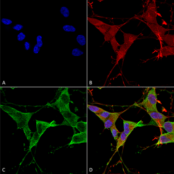
Immunocytochemistry/Immunofluorescence analysis using Mouse Anti-LAR/PTPRF Monoclonal Antibody, Clone N165/38. Tissue: Neuroblastoma cells (SH-SY5Y). Species: Human. Fixation: 4% PFA for 15 min. Primary Antibody: Mouse Anti-LAR/PTPRF Monoclonal Antibody at 1:100 for overnight at 4°C with slow rocking. Secondary Antibody: AlexaFluor 488 at 1:1000 for 1 hour at RT. Counterstain: Phalloidin-iFluor 647 (red) F-Actin stain; Hoechst (blue) nuclear stain at 1:800, 1.6mM for 20 min at RT. (A) Hoechst (blue) nuclear stain. (B) Phalloidin-iFluor 647 (red) F-Actin stain. (C) LAR/PTPRF Antibody (D) Composite.
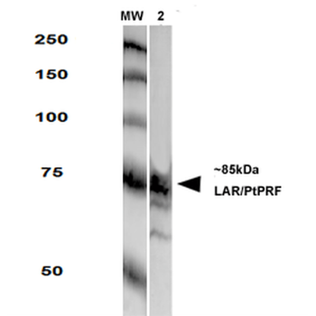
Western Blot analysis of Rat Brain Membrane showing detection of LAR protein using Mouse Anti-LAR Monoclonal Antibody, Clone N165/38. Primary Antibody: Mouse Anti-LAR Monoclonal Antibody at 1:250.
Quick Database Links
UniProt Details
− No UniProt data available
NCBI Gene Details
− No NCBI Gene data available
NCBI Reference Sequences
−Associated Accession Numbers
Curated reference sequences for the gene transcript and protein product| Protein | NP_002831.2 |
|---|
Documents Download
Datasheet
Product Information
Request a Document
Protocol Information
WB
Western Blot (IB, immunoblot)
IF
Immunofluorescence
ICC
Immunocytochemistry
LAR Antibody (PerCP) (orb167667)

- 0.0
Based on 0 reviews
Participating in our Biorbyt product reviews program enables you to support fellow scientists by sharing your firsthand experience with our products.
Login to Submit a ReviewAvailable Sizes
Select a size below
Choose Conjugation or Carrier Free Version
Free Secondary Antibody (20 ul)0/0
Please add an antibody product to your cart first.