You have no items in your shopping cart.
Featured
Description
Research Area
Cancer Biology, Neuroscience, Pharmacology & Drug Discovery
Images & Validation
−Item 1 of 2
| Tested Applications | ICC, IF, IHC, WB |
|---|---|
| Dilution Range | WB (1:1000), ICC/IF (1:100) |
| Reactivity | Human, Mouse, Rat |
| Application Notes |
Key Properties
−| Host | Mouse |
|---|---|
| Clonality | Monoclonal |
| Isotype | IgG2A |
| Clone No. | N366/60 (Formerly sold as S366-60) |
| Immunogen | Fusion protein amino acids 306-424 (Cytoplasmic C-terminus) of rat Kir6.1 |
| Target | Kir6.1 |
| Molecular Weight | 45kDa |
| Purification | Protein G Purified |
| Conjugation | Biotin |
Storage & Handling
−| Storage | Conjugated antibodies should be stored according to the product label |
|---|---|
| Buffer/Preservatives | 136.36mM Ethanolamine, 133.23 mM Chlorides, 9.55mM Phosphates, 9.55mM Sodium Bicarbonate |
| Concentration | 1 mg/ml |
| Expiration Date | 12 months from date of receipt. |
| Disclaimer | For research use only |
Alternative Names
−Kir6.1, KCNJ8, Kcnj8, Inward rectifier K(+) channel Kir6.1, Potassium channel inwardly rectifying subfamily J member 8, ATP-sensitive inward rectifier potassium channel 8, uKATP-1
Similar Products
−
Kir 6.1 Rabbit Polyclonal Antibody (Biotin) [orb459737]
WB
Bovine, Gallus, Human, Porcine, Rabbit
Mouse, Rat
Rabbit
Polyclonal
Biotin
100 μlKir6.1/KCNJ8 Rabbit Polyclonal Antibody (Biotin) [orb2603550]
ELISA, IHC, WB
Human, Mouse, Rat
Rabbit
Polyclonal
Biotin
100 μgKCNJ8 Rabbit Polyclonal Antibody (Biotin) [orb2133839]
WB
Bovine, Canine, Equine, Guinea pig, Human, Mouse, Rabbit, Rat, Zebrafish
Rabbit
Polyclonal
Biotin
100 μl
Quality Guarantee
Explore bioreagents carefree to elevate your research. All our products are rigorously tested for performance. If a product does not perform as described on its datasheet, our scientific support team will provide expert troubleshooting, a prompt replacement, or a refund. For full details, please see our Terms & Conditions and Buying Guide. Contact us at [email protected].

Immunocytochemistry/Immunofluorescence analysis using Mouse Anti-Kir6.1 Monoclonal Antibody, Clone N366/60. Tissue: Neuroblastoma cells (SH-SY5Y). Species: Human. Fixation: 4% PFA for 15 min. Primary Antibody: Mouse Anti-Kir6.1 Monoclonal Antibody at 1:100 for overnight at 4°C with slow rocking. Secondary Antibody: AlexaFluor 488 at 1:1000 for 1 hour at RT. Counterstain: Phalloidin-iFluor 647 (red) F-Actin stain; Hoechst (blue) nuclear stain at 1:800, 1.6mM for 20 min at RT. (A) Hoechst (blue) nuclear stain. (B) Phalloidin-iFluor 647 (red) F-Actin stain. (C) Kir6.1 Antibody (D) Composite.
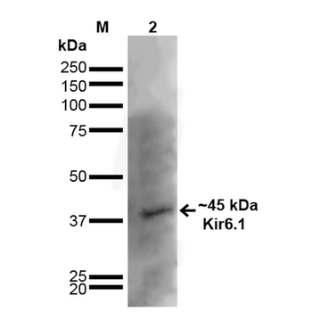
Western Blot analysis of Rat Brain showing detection of ~45 kDa Kir6.1 protein using Mouse Anti-Kir6.1 Monoclonal Antibody, Clone N366/60. Lane 1: MW Ladder. Lane 2: Rat Brain. Load: 20 μg. Block: 2% GE Healthcare Blocker for 1 hour at RT. Primary Antibody: Mouse Anti-Kir6.1 Monoclonal Antibody at 1:1000 for 16 hours at 4°C. Secondary Antibody: Goat Anti-Mouse IgG: HRP at 1:200 for 1 hour at RT. Color Development: ECL solution for 6 min at RT. Predicted/Observed Size: ~45 kDa. Other Band (s): ~100 kDa.
Quick Database Links
UniProt Details
− No UniProt data available
NCBI Gene Details
− No NCBI Gene data available
NCBI Reference Sequences
−Associated Accession Numbers
Curated reference sequences for the gene transcript and protein product| Protein | NP_058795.3 |
|---|
Documents Download
Datasheet
Product Information
Request a Document
Protocol Information
WB
Western Blot (IB, immunoblot)
IHC
Immunohistochemistry
IF
Immunofluorescence
ICC
Immunocytochemistry
Kir6.1 Antibody (Biotin) (orb376929)

- 0.0
Based on 0 reviews
Participating in our Biorbyt product reviews program enables you to support fellow scientists by sharing your firsthand experience with our products.
Login to Submit a ReviewAvailable Sizes
Select a size below
Choose Conjugation or Carrier Free Version
Free Secondary Antibody (20 ul)0/0
Please add an antibody product to your cart first.