You have no items in your shopping cart.
Description
Research Area
Neuroscience
Images & Validation
−Item 1 of 4
| Tested Applications | ELISA, IF, IHC, WB |
|---|---|
| Dilution Range | ELISA: 1:20,000 - 1:100,000, IHC: 1:100 - 1:500, IF: 1:200 - 1:1,000, WB: 1:500 - 1:2,000 |
| Reactivity | Human, Mouse |
| Application Notes |
Key Properties
−| Antibody Type | Primary Antibody |
|---|---|
| Host | Rabbit |
| Clonality | Polyclonal |
| Isotype | IgG |
| Immunogen | This protein A purified Jagged 1 antibody was prepared from whole rabbit serum produced by repeated immunizations with a synthetic peptide corresponding to amino acids near the N-terminus of human Jagged-1 protein. |
| Target | JAG1 |
| Purity | This protein A purified antibody is directed against human Jagged-1 protein. The product was purified from mono-specific antiserum by affinity chromatography. A BLAST analysis was used to suggest cross reactivity with Jagged-1 protein from human, chimpanzee, rat and mouse based on 100% homology with the immunizing sequence. Partial reactivity is expected against dog (81%) and Xenopus laevis (85%) based on partial sequence homologies as indicated. Reactivity against homologues from other sources is not known. |
| Conjugation | Unconjugated |
Storage & Handling
−| Storage | Store vial at -20° C or below prior to opening. This vial contains a relatively low volume of reagent (25 µL). To minimize loss of volume dilute 1:10 by adding 225 µL of the buffer stated above directly to the vial. Recap, mix thoroughly and briefly centrifuge to collect the volume at the bottom of the vial. Use this intermediate dilution when calculating final dilutions as recommended below. Store the vial at -20°C or below after dilution. Avoid cycles of freezing and thawing. |
|---|---|
| Form/Appearance | Liquid (sterile filtered) |
| Buffer/Preservatives | Preservative: 0.01% (w/v) Sodium Azide. Stabilizer: None; Buffer: 0.02 M Potassium Phosphate, 0.15 M Sodium Chloride, pH 7.2 |
| Concentration | 1.1 mg/mL |
| Expiration Date | 12 months from date of receipt. |
| Dry Ice Shipping | Please note: This product requires shipment on dry ice. A dry ice surcharge will apply. |
| Disclaimer | For research use only |
Alternative Names
−rabbit anti-Jagged 1 Antibody, rabbit anti-Jagged1 Antibody, rabbit anti-Jagged-1 Antibody, Ser 1 antibody, AGS antibody, AHD antibody, AWS antibody, CD 339 antibody, CD339 antibody, CD339 antigen antibody, Headturner antibody, HJ1 antibody, Htu antibody
Similar Products
−- Item 1 of 4
Jagged1 Rabbit Polyclonal Antibody [orb10065]
ICC, IF, IHC-Fr, IHC-P, WB
Mouse, Rat
Human
Rabbit
Polyclonal
Unconjugated
50 μl, 100 μl, 200 μl - Item 1 of 7
Jagged1 Polyclonal Antibody [orb1411370]
IF, IHC-P, WB
Human, Mouse, Rat
Rabbit
Polyclonal
Unconjugated
100 μl - Item 1 of 4
Jagged1 rabbit pAb Antibody [orb767006]
ELISA, IF, IHC, WB
Human, Mouse, Rat
Polyclonal
Unconjugated
100 μl, 50 μl - Item 1 of 1
- Item 1 of 1


Quality Guarantee
Explore bioreagents carefree to elevate your research. All our products are rigorously tested for performance. If a product does not perform as described on its datasheet, our scientific support team will provide expert troubleshooting, a prompt replacement, or a refund. For full details, please see our Terms & Conditions and Buying Guide. Contact us at [email protected].
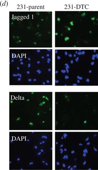
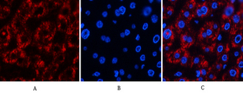
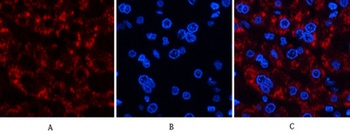
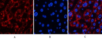
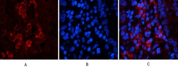
(a) Representative confocal microscopy shows CD44, CD24 and cleaved notch (NICD) in a population of drug naive MDA-MB-231. Yellow arrows indicate CD44HiCD24Lo (M) population of cells and the white arrows indicate the CD44HiCD24Hi (E/M) cells. Histogram (right panel) shows quantification of NICD in the distinct phenotype populations (M versus E/M). N = 3 biological replicates. (b) Schematic describes the experimental protocol to generate drug-tolerant cells (DTCs) parental MDA-MB-231 cells were treated with docetaxel at 100 nM (20× the IC50) and subsequently selected by substrate re-attachment and acute population outgrowth. (c) Representative confocal microscopy shows CD44, CD24 and NICD in the MDA-MB-231 parent and DTC populations. Right panel shows quantification of fluorescence intensity of each signal determined by at least 25 individual fields. N = 3 biological replicates. (d) Representative confocal microscopy shows Jagged and Delta expression in MDA-MB-231 parent and DTC. DAPI nuclear stain (blue). N = 3 biological replicates.
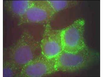
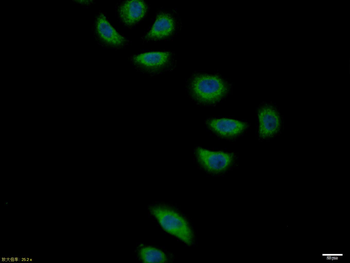
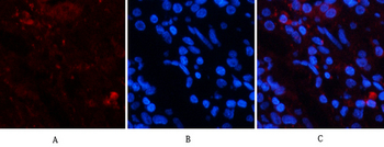
Immunofluorescence microscopy using Biorbyt's Protein A purified anti-Jagged-1 antibody of human corneal epithelial cells. Primary antibody was used at a 1:500 dilution. The Jagged1 (green staining) is localized to the cytoplasm and is consistent with reports in the literature. The nucleus is stained with Bis benzamine (blue).
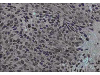
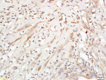
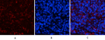
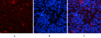
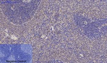
Immunohistochemical staining of human cervical cancer tissue (40X magnification) using Biorbyt's Protein A purified anti-Jagged-1 antibody. Tissue was fixed with formalin and embedded in paraffin. Hematoxylin was used to counter-stain cells. A 1:100 dilution of primary antibody was used.
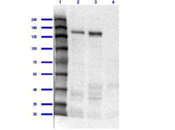
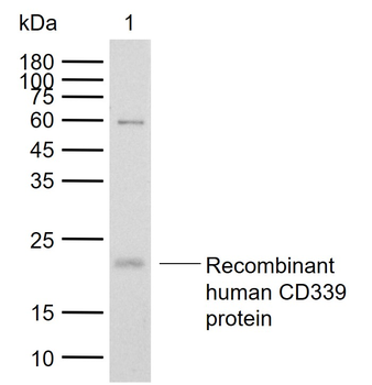
Western Blot of Rabbit Anti-Jagged 1 Antibody. Lane 1: Opal Prestained MW marker. Lane 2: Mouse Liver Whole Cell Lysate [10 µg] (p/n orb348718). Lane 3: Human Liver Whole Cell Lysate [10 µg]. Lane 4: Human Lung Whole Cell Lysate [10 µg]. Primary Antibody: Anti-Jagged 1 at 1:1000 overnight at 2-8°C. Secondary Antibody: Goat Anti-Rabbit IgG Peroxidase Conjugated (p/n orb347654) at 1:70000 for 30 mins at RT. Blocking Buffer: BlockOut Buffer (p/n orb348644) for 1hr RT. Predicted Molecular Weight: 113kDa. Exposure: 30 sec.
Documents Download
Datasheet
Product Information
Request a Document
Protocol Information
WB
Western Blot (IB, immunoblot)
IHC
Immunohistochemistry
IF
Immunofluorescence
ELISA
Enzyme-linked Immunosorbent Assay (EIA)
JAG1 Antibody (orb344625)

- 0.0
Based on 0 reviews
Participating in our Biorbyt product reviews program enables you to support fellow scientists by sharing your firsthand experience with our products.
Login to Submit a ReviewAvailable Sizes
Select a size below
Free Secondary Antibody (20 ul)0/0
Please add an antibody product to your cart first.