You have no items in your shopping cart.
Description
Research Area
Immunology & Inflammation
Images & Validation
−Item 1 of 3
| Tested Applications | ELISA, IF, IHC, WB |
|---|---|
| Dilution Range | ELISA: 1:1,000 - 1:5,000, IF: 1:50-1:200, WB: 1:500 - 1:2,000 |
| Reactivity | Mouse |
| Application Notes |
Key Properties
−| Antibody Type | Primary Antibody |
|---|---|
| Host | Rabbit |
| Clonality | Polyclonal |
| Isotype | IgG |
| Immunogen | The whole rabbit serum used to produce this IgG fraction antibody was prepared by repeated immunizations with native 157 aa mouse IL-18 produced in E.coli. |
| Target | Il18 |
| Purity | This is an IgG preparation of whole rabbit serum purified by protein A chromatography using a low endotoxin methodology. This antibody is primarily directed against mature 18,000 MW mouse IL-18 and is useful in determining its presence in various assays. This antibody will also recognize the 24,000 inactive precursor form of mouse IL-18. In general, this antibody also detects rat IL-18 in the same formats using similar dilutions. A control of similarly diluted LOW ENDOTOXIN CONTROL RABBIT IgG is recommended. |
| Conjugation | Unconjugated |
Storage & Handling
−| Storage | Store vial at -20° C or below prior to opening. This vial contains a relatively low volume of reagent (25 µL). To minimize loss of volume dilute 1:10 by adding 225 µL of the buffer stated above directly to the vial. Recap, mix thoroughly and briefly centrifuge to collect the volume at the bottom of the vial. Use this intermediate dilution when calculating final dilutions as recommended below. Store the vial at -20°C or below after dilution. Avoid cycles of freezing and thawing. |
|---|---|
| Form/Appearance | Liquid (sterile filtered) |
| Buffer/Preservatives | Preservative: None. Stabilizer: None; Buffer: 0.02 M Potassium Phosphate, 0.15 M Sodium Chloride, pH 7.2 |
| Concentration | 1.0 mg/ml |
| Expiration Date | 12 months from date of receipt. |
| Dry Ice Shipping | Please note: This product requires shipment on dry ice. A dry ice surcharge will apply. |
| Disclaimer | For research use only |
Alternative Names
−rabbit anti-IL-18 antibody, rabbit anti-interleukin-18 antibody, Iboctadekin antibody, IFN gamma inducing factor antibody, IGIF antibody, IL 1 gamma antibody, IL 18 antibody, IL 1g antibody, IL-1F4, IL 18, Interleukin 18, IL18, Interleukin18, IL1 F4, IL1F4, Ms IL-18, mouse IL18
Similar Products
−- Item 1 of 6
IL18 Antibody (C-term) [orb1927175]
FC, IF, IHC-P, WB
Human
Rabbit
Polyclonal
Unconjugated
50 μl, 100 μl - Item 1 of 5
IL18/IL1F4 Human Monoclonal Antibody [orb2960417]
ELISA, FC, NeA
Human
Human
Monoclonal
Unconjugated
1 mg, 100 μg - Item 1 of 4
IL18 Rabbit Polyclonal Antibody [orb10899]
WB
Human
Human
Rabbit
Polyclonal
Unconjugated
50 μl, 100 μl, 200 μl - Item 1 of 1
- Item 1 of 4
IL18 Antibody [orb2309942]
IHC
Canine, Equine, Feline, Human
Mouse
Monoclonal
Unconjugated
100 μg, 20 μg, 100 μg (without BSA and Azide)


Quality Guarantee
Explore bioreagents carefree to elevate your research. All our products are rigorously tested for performance. If a product does not perform as described on its datasheet, our scientific support team will provide expert troubleshooting, a prompt replacement, or a refund. For full details, please see our Terms & Conditions and Buying Guide. Contact us at [email protected].
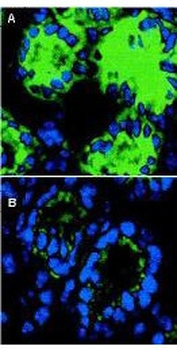
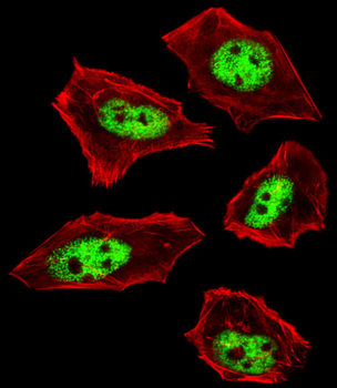
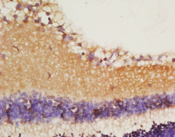
Immunofluorescence microscopy of IL-18 in mouse colon sections. The transversing portion of the large intestine from DSS-exposed (Panel A) and -unexposed mice (Panel B) was excised, rinsed in PBS, and frozen on isopentane cooled with liquid nitrogen. Frozen sections (5 µm) were cut on a Leica CM 1850 cryostat. The slides were fixed for 10 min in 4% paraformaldehyde, air-dried, and incubated for 20 min in PBS supplemented with 10% normal goat serum. Sections were incubated in a 1:50 dilution of Biorbyt's rabbit anti-Mouse IL-18 antibody or 1 µg/ml nonimmune rabbit IgG (not shown) as negative control. The antibodies were diluted in PBS containing 1% bovine serum albumin. After an overnight incubation at 4°C, the sections were washed three times with 0.5% bovine serum albumin in PBS. The sections were then incubated with a secondary goat anti-rabbit antibody conjugated to Alexa488 (Molecular Probes) for 60 min at room temperature in the dark. Nuclei were counterstained blue using 1 µg/100 ml bisbenzimide. After staining, sections were washed and examined with the Leica DM RXA confocal laser scanning system and analyzed. Similar staining will occur with other systems.
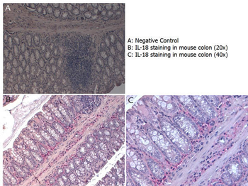
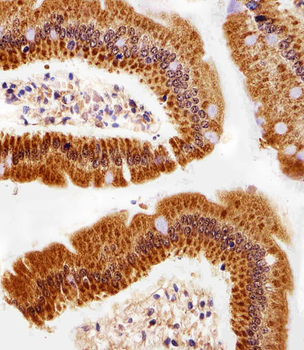
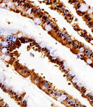
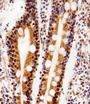
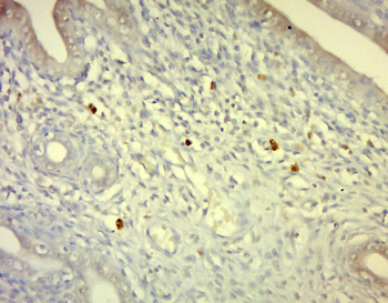
Immunohistochemistry with Rabbit anti-Mouse IL-18 antibody showing IL-18 staining in inflammatory cells of the mucous corium of mouse colon at 20x and 40x. Slide A is a negative control. Slides B and C show staining. Formalin fixed/paraffin embedded sections were subjected to heat induced epitope retrieval (HIER) at pH6.2 and then incubated with mouse anti-IL-18 antibody at 4.0 µg/ml for 60 minutes. The reaction was developed using MACH 4 universal AP polymer detection system and visualized with WARP RED.
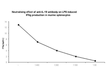
In-vitro neutralization. Spleens were aseptically removed and cell suspensions were prepared. Cells were washed twice and resuspended in RPMI supplemented with 10% FBS. For cytokine measurement, spleen cells were cultured at 5 mln/mL in 24-well, flat-bottom culture plates in the presence of several dilutions of rabbit anti-murine IL-18 antibody (1:400; 1:200; 1:100; 1:50) and 100 ng/mL of LPS. Cultures were incubated at 37°C in a humidified atmosphere with 5% CO2. At the end of the incubation period, cultures were frozen at -70°C and subjected to 3 freeze-thaw cycles to obtain total cytokine levels. Before assaying, samples were centrifuged for 10 minutes at 10000g to remove debris.
Documents Download
Datasheet
Product Information
Request a Document
Protocol Information
WB
Western Blot (IB, immunoblot)
IHC
Immunohistochemistry
IF
Immunofluorescence
ELISA
Enzyme-linked Immunosorbent Assay (EIA)
Il18 Antibody (orb345221)

- 0.0
Based on 0 reviews
Participating in our Biorbyt product reviews program enables you to support fellow scientists by sharing your firsthand experience with our products.
Login to Submit a ReviewAvailable Sizes
Select a size below
Free Secondary Antibody (20 ul)0/0
Please add an antibody product to your cart first.