You have no items in your shopping cart.
Description
Research Area
Immunology & Inflammation
Images & Validation
−Item 1 of 1
| Tested Applications | ELISA, IHC, WB |
|---|---|
| Dilution Range | ELISA: 1:20,000-1:100,000, IHC: 1:1,000-1:5,000, WB: 1:2,000-1:10,000 |
| Reactivity | Human |
| Application Notes |
Key Properties
−| Antibody Type | Primary Antibody |
|---|---|
| Host | Rabbit |
| Clonality | Polyclonal |
| Isotype | IgG |
| Immunogen | This purified antibody was prepared from whole rabbit serum produced by repeated immunizations with full length recombinant human IL17-A protein. |
| Target | IL17A |
| Purity | This purified antibody has been heated to 56°C for 30 minutes. In ELISA and other immunoreactive assays, this antibody will recognize both native and recombinant human IL17-A in cell supernatants and certain body fluids. A control of similarly diluted normal rabbit IgG is recommended. |
| Conjugation | Biotin |
Storage & Handling
−| Storage | Store vial at 4° C prior to restoration. For extended storage aliquot contents and freeze at -20° C or below. Avoid cycles of freezing and thawing. Centrifuge product if not completely clear after standing at room temperature. This product is stable for several weeks at 4° C as an undiluted liquid. Dilute only prior to immediate use. |
|---|---|
| Form/Appearance | Lyophilized |
| Buffer/Preservatives | Preservative: 0.01% (w/v) Sodium Azide. Stabilizer: 10 mg/mL Bovine Serum Albumin (rAlbumin) - Immunoglobulin and Protease free; Buffer: 0.02 M Potassium Phosphate, 0.15 M Sodium Chloride, pH 7.2 |
| Concentration | 1.0 mg/ml |
| Expiration Date | 12 months from date of receipt. |
| Disclaimer | For research use only |
Alternative Names
−rabbit anti-Interleukin-17A biotin conjugated antibody, rabbit anti-IL-17A biotin conjugated antibody, IL-17A, Interleukin-17A, Cytotoxic T-Lymphocyte-associated Antigen 8, CTLA8, Interleukin-17, IL17, IL-17, Interleukin17
Similar Products
−- Item 1 of 1

Quality Guarantee
Explore bioreagents carefree to elevate your research. All our products are rigorously tested for performance. If a product does not perform as described on its datasheet, our scientific support team will provide expert troubleshooting, a prompt replacement, or a refund. For full details, please see our Terms & Conditions and Buying Guide. Contact us at [email protected].
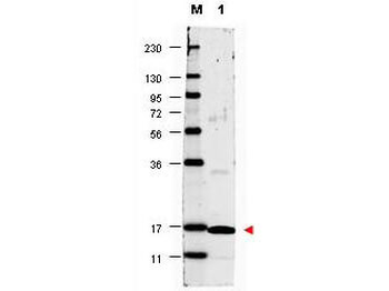
Western blot using Biorbyt's anti-Human IL17-A antibody shows detection of a band ~17 kDa in size corresponding to recombinant human IL17-A (lane 1). Molecular weight markers are also shown (M). After transfer, the membrane was blocked overnight with 3% BSA in TBS followed by reaction with primary antibody at a 1:1000 dilution. Detection occurred using DyLight 649 conjugated anti-Rabbit IgG secondary antibody diluted 1:20000 in blocking buffer (p/n orb348637).
Quick Database Links
UniProt Details
− No UniProt data available
NCBI Reference Sequences
−Associated Accession Numbers
Curated reference sequences for the gene transcript and protein product| RefSeq | AAH662531.1 |
|---|
Documents Download
Datasheet
Product Information
Request a Document
Protocol Information
WB
Western Blot (IB, immunoblot)
IHC
Immunohistochemistry
ELISA
Enzyme-linked Immunosorbent Assay (EIA)
IL17A Antibody (Biotin) (orb345174)

- 0.0
Based on 0 reviews
Participating in our Biorbyt product reviews program enables you to support fellow scientists by sharing your firsthand experience with our products.
Login to Submit a ReviewAvailable Sizes
Select a size below
Choose Conjugation or Carrier Free Version
Free Secondary Antibody (20 ul)0/0
Please add an antibody product to your cart first.