You have no items in your shopping cart.
Description
Research Area
Metabolism Research
Images & Validation
−Item 1 of 7
| Tested Applications | FC, IF, IHC-P, WB |
|---|---|
| Dilution Range | IHC-P - 1:100-500, WB - 1:1000, IF - 1:10-50, FC - 1:10-50 |
| Reactivity | Human, Mouse, Rat |
| Predicted Reactivity | Bovine |
Key Properties
−| Antibody Type | Primary Antibody |
|---|---|
| Host | Rabbit |
| Clonality | Polyclonal |
| Isotype | Rabbit IgG |
| Molecular Weight | 46659 Da |
| Conjugation | Unconjugated |
Storage & Handling
−| Storage | Maintain refrigerated at 2-8°C for up to 2 weeks. For long term storage store at -20°C in small aliquots to prevent freeze-thaw cycles |
|---|---|
| Form/Appearance | Purified polyclonal antibody supplied in PBS with 0.09% (W/V) sodium azide. This antibody is prepared by Saturated Ammonium Sulfate (SAS) precipitation followed by dialysis against PBS. |
| Expiration Date | 12 months from date of receipt. |
| Disclaimer | For research use only |
Alternative Names
−PICD
Similar Products
−- Item 1 of 5
IDH1 Antibody (Center) [orb1788320]
FC, IF, IHC-P, WB
Mouse, Sheep
Human, Rat
Rabbit
Polyclonal
Unconjugated

Quality Guarantee
Explore bioreagents carefree to elevate your research. All our products are rigorously tested for performance. If a product does not perform as described on its datasheet, our scientific support team will provide expert troubleshooting, a prompt replacement, or a refund. For full details, please see our Terms & Conditions and Buying Guide. Contact us at [email protected].
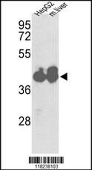
Western blot analysis of IDH1 Antibody (Center) in HepG2 cell line and mouse liver tissue lysates (35 ug/lane). IDH1 (arrow) was detected using the purified Pab.
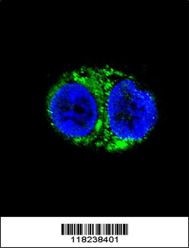
Confocal immunofluorescent analysis of IDH1 Antibody (Center) with HepG2 cell followed by Alexa Fluor 488-conjugated goat anti-rabbit lgG (green).DAPI was used to stain the cell nuclear (blue).
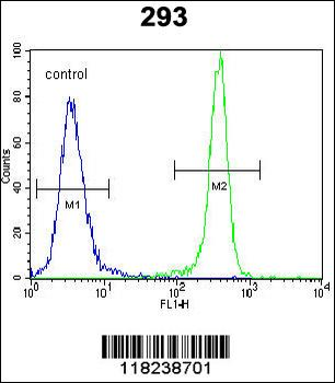
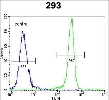
IDH1 Antibody (Center) flow cytometric analysis of 293 cells (right histogram) compared to a negative control cell (left histogram). FITC-conjugated goat-anti-rabbit secondary antibodies were used for the analysis.
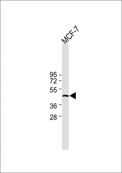
All lanes: Anti-IDH1 Antibody (Center) at 1:2000 dilution. Lane 1: MCF-7 whole cell lysate. Lysates/proteins at 20 µg per lane. Secondary Goat Anti-Mouse IgG/A/M (H/L), Peroxidase conjugated at 1/2000 dilution.Observed band size: 47kDa. Blocking/Dilution buffer: 5% NFDM/TBST.

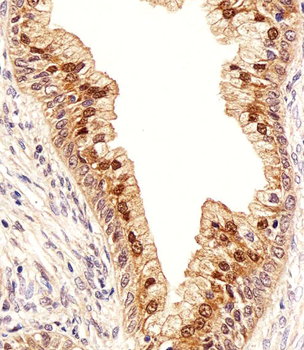
Immunohistochemical analysis of paraffin-embedded H. prostate section using IDH1 Antibody (Center). Diluted at 1:100 dilution. A peroxidase-conjugated goat anti-rabbit IgG at 1:400 dilution was used as the secondary antibody, followed by DAB staining.
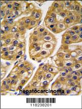
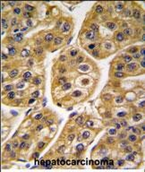
Formalin-fixed and paraffin-embedded human hepatocarcinoma tissue reacted with IDH1 antibody (Center), which was peroxidase-conjugated to the secondary antibody, followed by DAB staining. This data demonstrates the use of this antibody for immunohistochemistry; clinical relevance has not been evaluated.
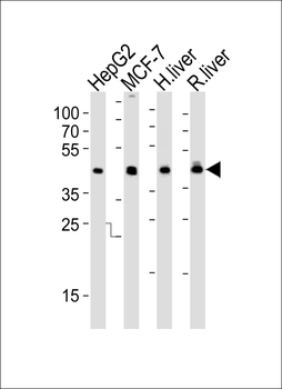
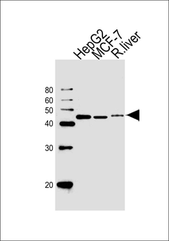
Western blot analysis of lysates from HepG2, MCF-7 cell line, human liver and rat liver tissue lysate (from left to right), using IDH1 Antibody (Center). Diluted at 1:1000 at each lane. A goat anti-rabbit IgG H&L (HRP) at 1:10000 dilution was used as the secondary antibody. Lysates at 35 ug per lane.
Quick Database Links
UniProt
UniProt Details
− No UniProt data available
Documents Download
Datasheet
Product Information
Request a Document
Protocol Information
WB
Western Blot (IB, immunoblot)
IHC-P
Immunohistochemistry Paraffin
FC
Flow Cytometry
IF
Immunofluorescence
IDH1 Antibody (Center) (orb1929244)

- 0.0
Based on 0 reviews
Participating in our Biorbyt product reviews program enables you to support fellow scientists by sharing your firsthand experience with our products.
Login to Submit a ReviewAvailable Sizes
Select a size below
Free Secondary Antibody (20 ul)0/0
Please add an antibody product to your cart first.