You have no items in your shopping cart.
HPD Antibody
SKU: orb1262616
Featured
Description
Research Area
Cell Biology, Metabolism Research, Signal Transduction
Images & Validation
−Item 1 of 4
| Tested Applications | FC, IHC-P, WB |
|---|---|
| Reactivity | Human, Mouse |
| Application Notes |
Key Properties
−| Antibody Type | Primary Antibody |
|---|---|
| Host | Rabbit |
| Clonality | Polyclonal |
| Isotype | Rabbit Ig |
| Immunogen | This HPD antibody is generated from rabbits immunized with a KLH conjugated synthetic peptide between 92-121 amino acids from the N-terminal region of human HPD. |
| Target | HPD |
| Molecular Weight | 45 kDa |
| Purification | This antibody is purified through a protein A column, followed by peptide affinity purification. |
| Conjugation | Unconjugated |
Storage & Handling
−| Storage | Maintain refrigerated at 2-8°C for up to 2 weeks. For long term storage store at -20°C in small aliquots to prevent freeze-thaw cycles. |
|---|---|
| Form/Appearance | Liquid |
| Buffer/Preservatives | Supplied in PBS with 0.09% (W/V) sodium azide. |
| Concentration | batch dependent |
| Expiration Date | 12 months from date of receipt. |
| Disclaimer | For research use only |
Alternative Names
−4-hydroxyphenylpyruvate dioxygenase, 4-hydroxyphenylpyruvic acid oxidase, 4HPPD, HPD, HPPDase, HPD, PPD
Similar Products
−- Item 1 of 5
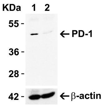
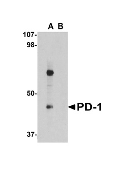
PD-1 Rabbit Polyclonal Antibody [orb13641]
FC
Human, Rat
Mouse
Rabbit
Polyclonal
Unconjugated
50 μl, 100 μl, 200 μl - Item 1 of 8
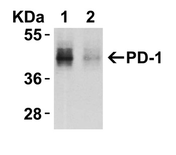
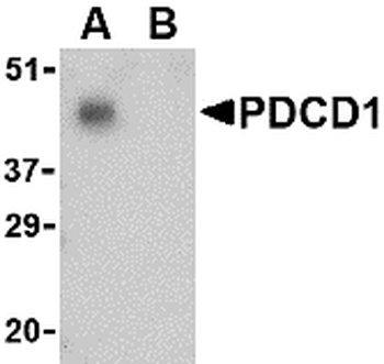
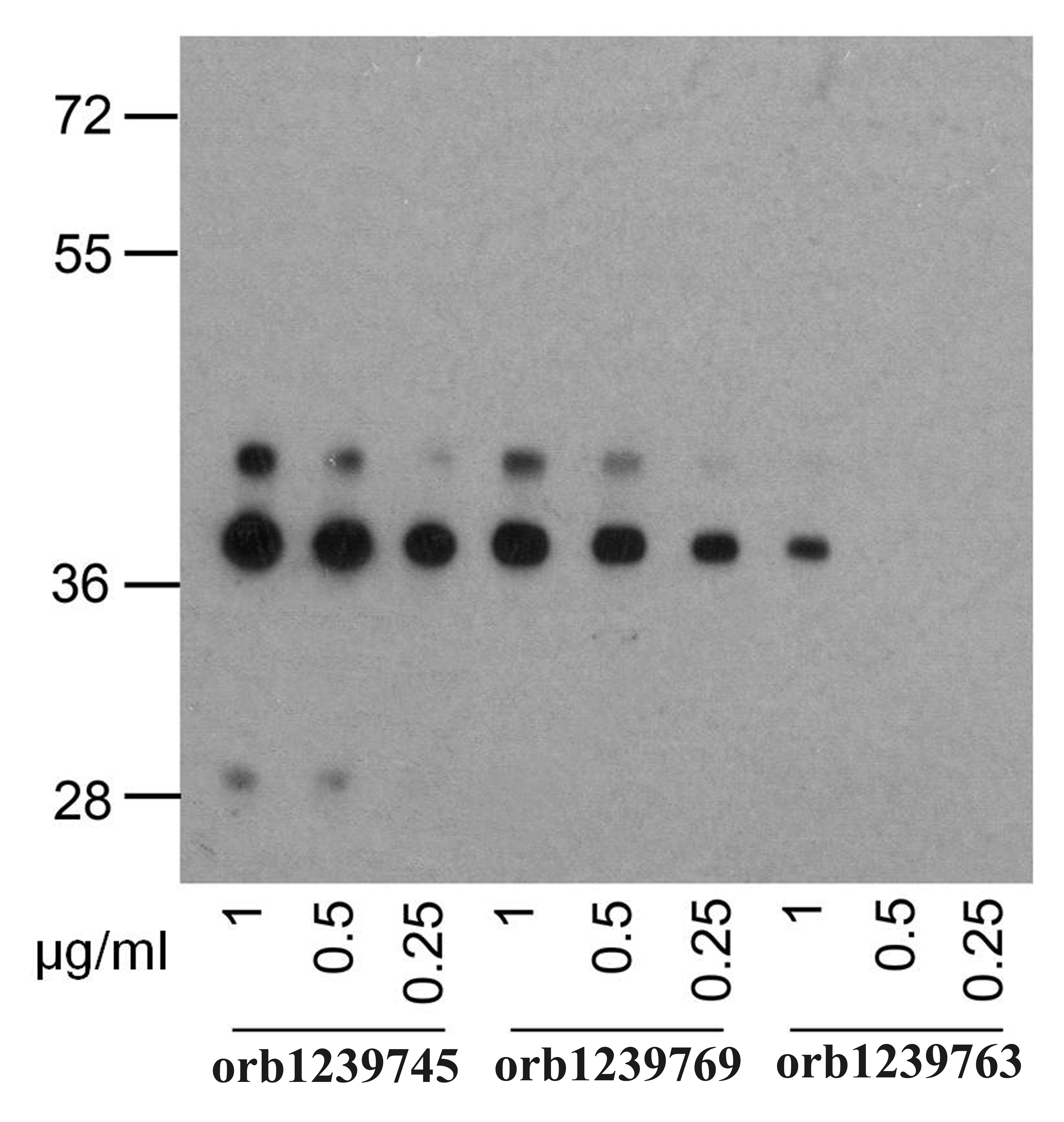
PDCD1 Antibody [orb1239763]
ELISA, FC, ICC, IF, IHC-P, WB
Human
Mouse
Monoclonal
Unconjugated
0.02 mg, 0.1 mg - Item 1 of 8
PDCD1 Antibody [orb1239732]
ELISA, IF, IHC-P, WB
Human, Mouse, Rat
Rabbit
Polyclonal
Unconjugated
0.1 mg, 0.02 mg - Item 1 of 7
PDCD1 Antibody [orb1239769]
ELISA, ICC, IF, IHC-P, WB
Human
Mouse
Monoclonal
Unconjugated
0.02 mg, 0.1 mg - Item 1 of 6
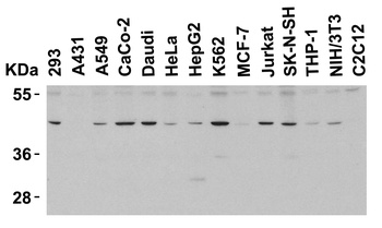
PD-1/PDCD1/PD Rabbit Polyclonal Antibody [orb1145869]
FC, IF, IHC, WB
Mouse, Rat
Rabbit
Polyclonal
Unconjugated
100 μg


Quality Guarantee
Explore bioreagents carefree to elevate your research. All our products are rigorously tested for performance. If a product does not perform as described on its datasheet, our scientific support team will provide expert troubleshooting, a prompt replacement, or a refund. For full details, please see our Terms & Conditions and Buying Guide. Contact us at [email protected].
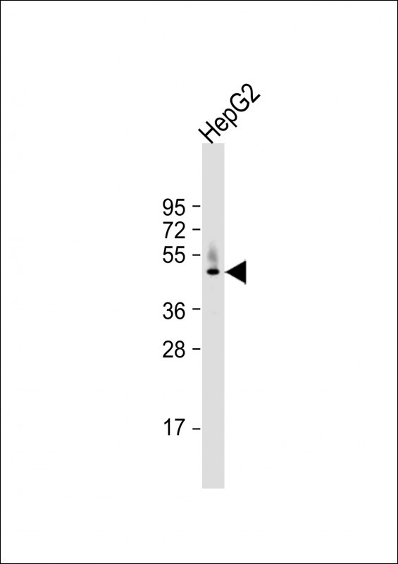
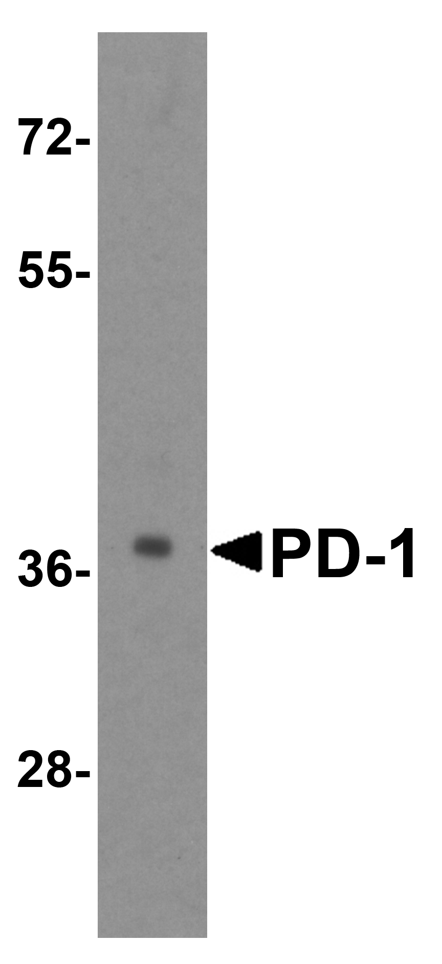
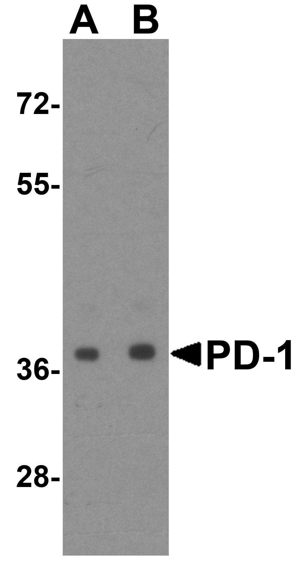
Western Blot at 1:1000 dilution + HepG2 whole cell lysate Lysates/proteins at 20 ug per lane.
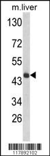
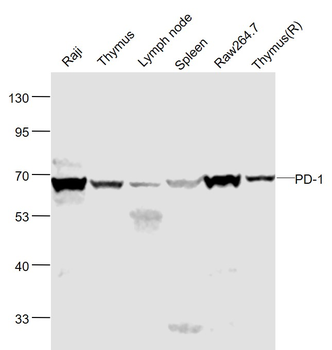
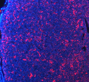
Western blot analysis of HPD Antibody in mouse liver tissue lysates (35 ug/lane)
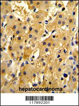
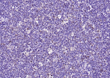
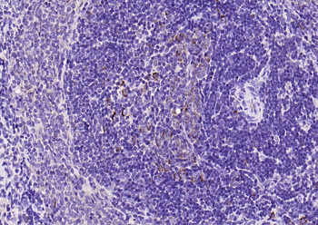
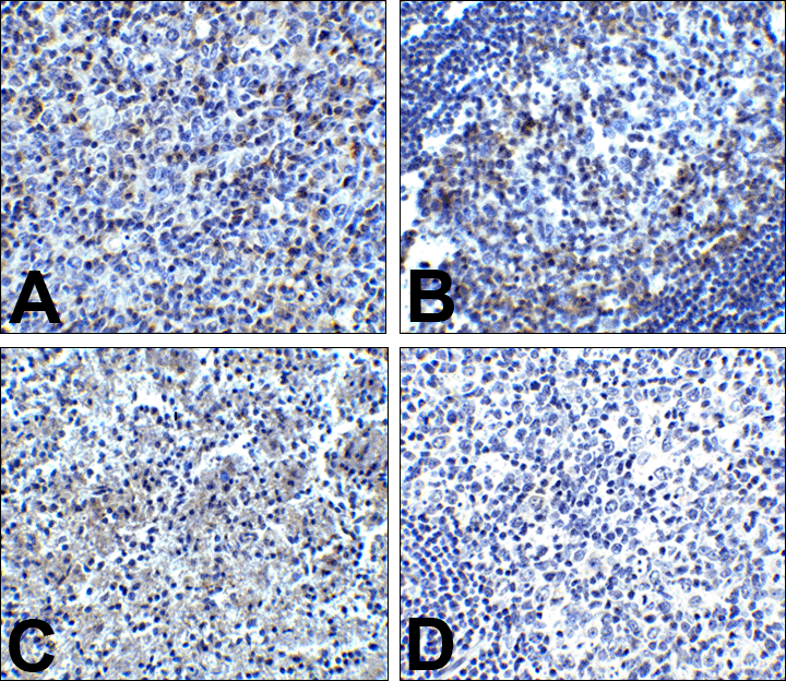
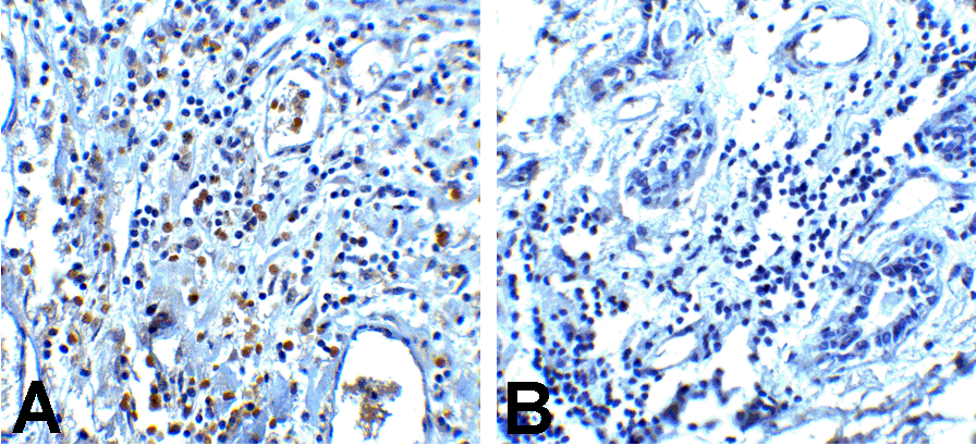
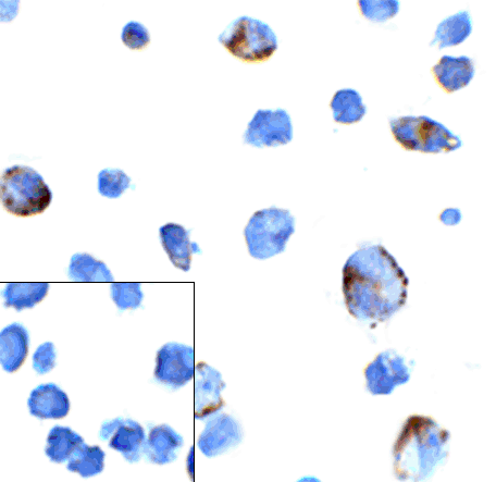
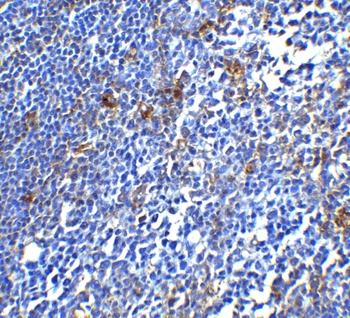
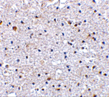
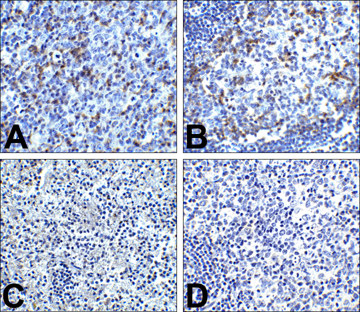
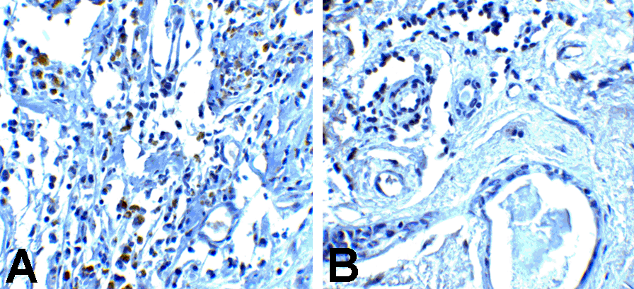
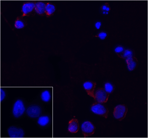
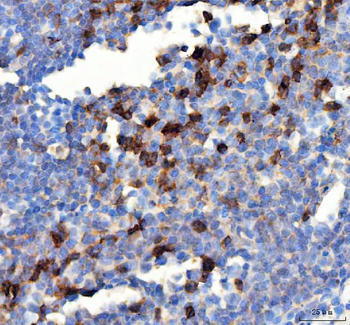
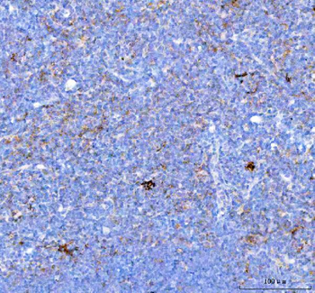
Formalin-fixed and paraffin-embedded human hepatocarcinoma reacted with HPD Antibody (N-term), which was peroxidase-conjugated to the secondary antibody, followed by DAB staining.
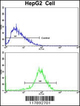
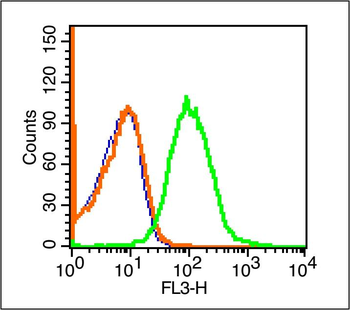
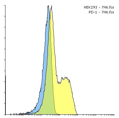
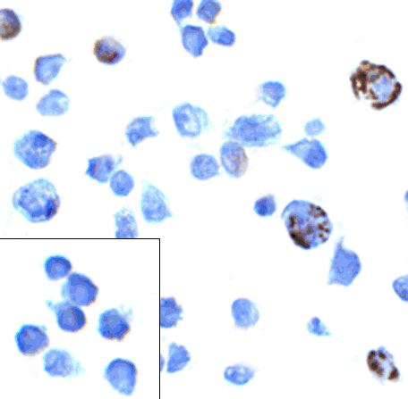
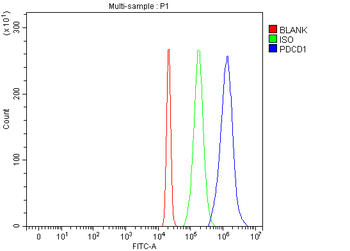
Flow cytometric analysis of HepG2 cells using HPD Antibody (N-term) (bottom histogram) compared to a negative control cell (top histogram). FITC-conjugated goat-anti-rabbit secondary antibodies were used for the analysis.
Documents Download
Datasheet
Product Information
Request a Document
Protocol Information
WB
Western Blot (IB, immunoblot)
IHC-P
Immunohistochemistry Paraffin
FC
Flow Cytometry
HPD Antibody (orb1262616)

- 0.0
Based on 0 reviews
Participating in our Biorbyt product reviews program enables you to support fellow scientists by sharing your firsthand experience with our products.
Login to Submit a ReviewAvailable Sizes
Select a size below