You have no items in your shopping cart.
Featured
Description
Research Area
Cell Biology
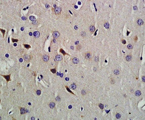
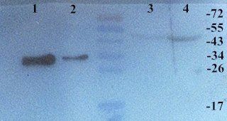
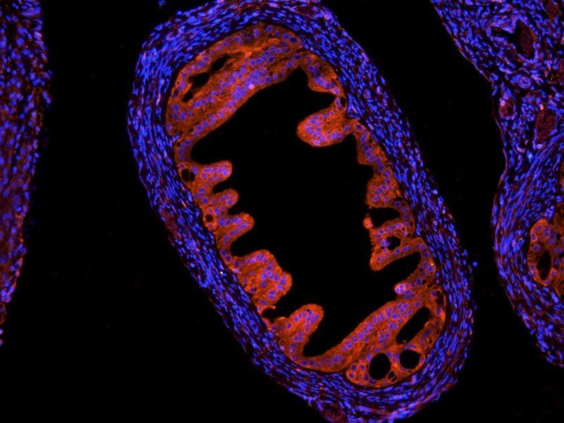
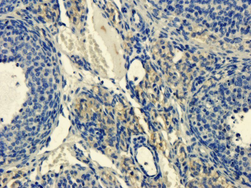
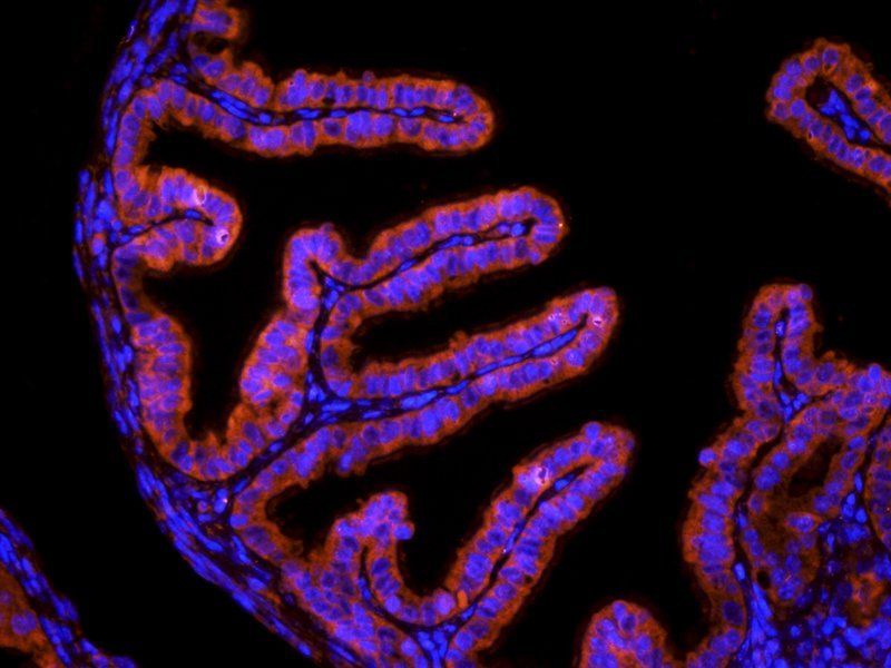
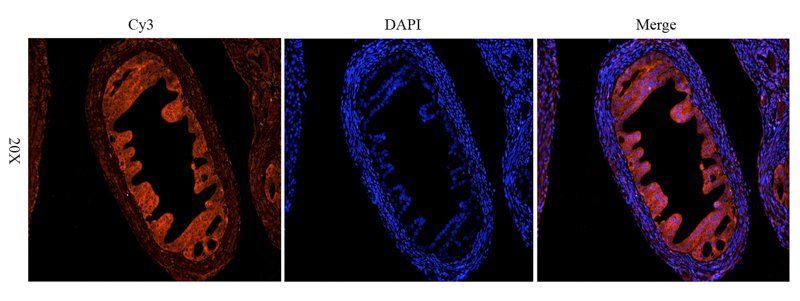
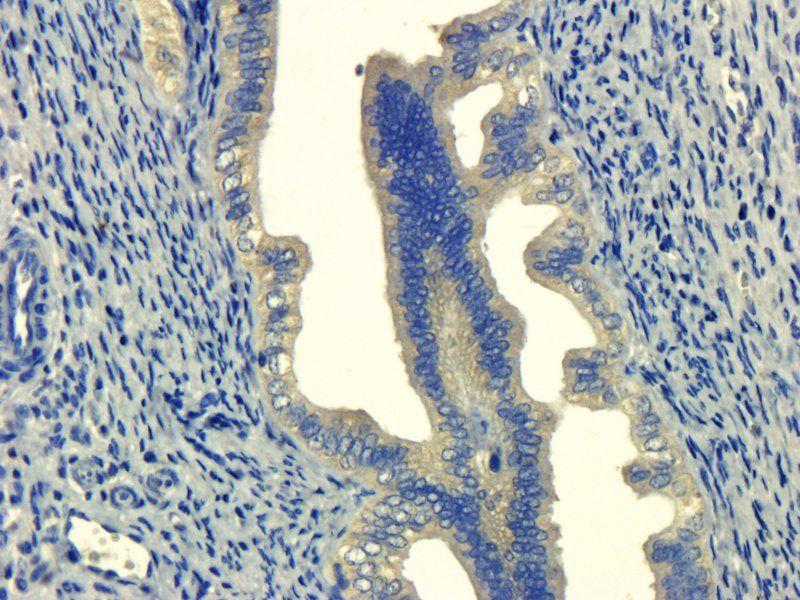
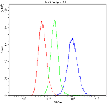
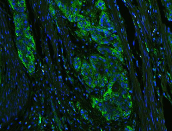
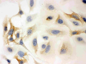
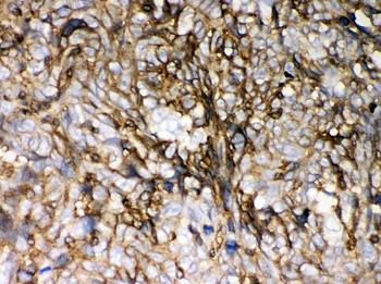
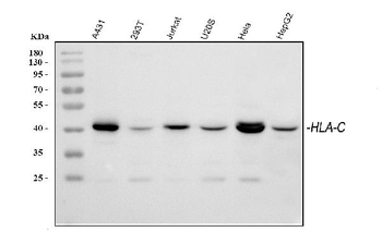
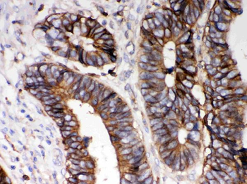
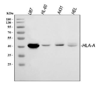
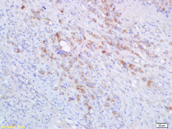
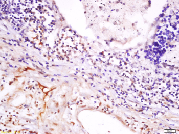
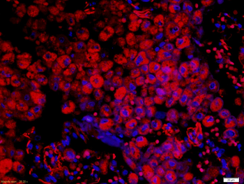
Images & Validation
−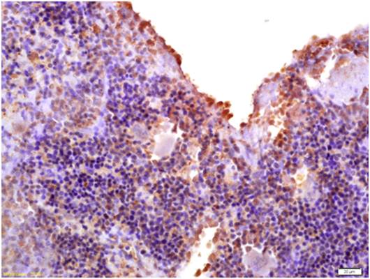
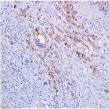
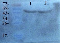
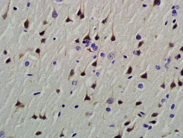
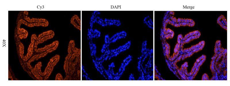
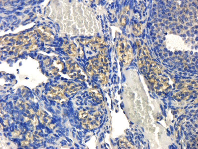
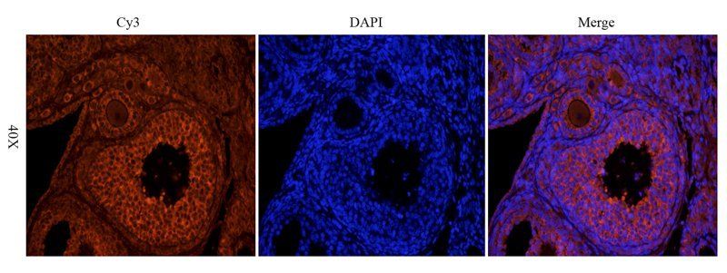
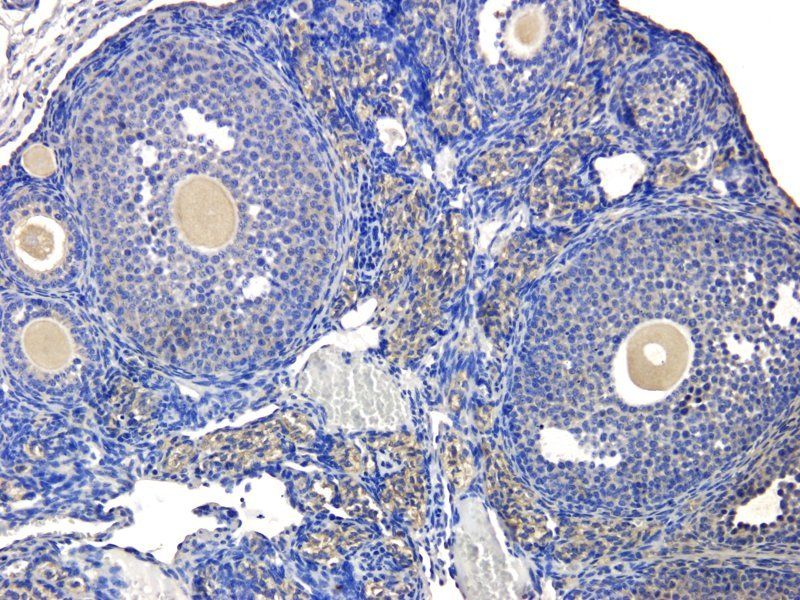
Item 1 of 15
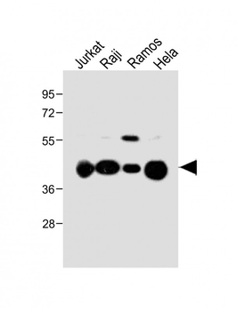
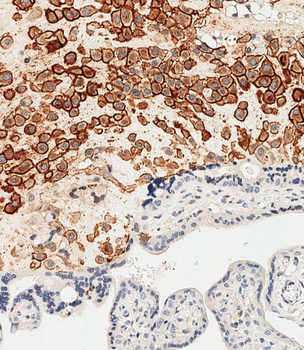
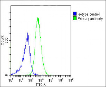
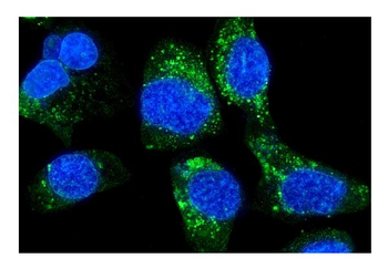
| Tested Applications | ELISA, ICC, IF, IHC-P, WB |
|---|---|
| Dilution Range | WB: 2 μg/ml, IF/ICC: 1:100, IHC-P: 1:100 |
| Reactivity | Human |
| Application Notes |
Key Properties
−| Host | Rabbit |
|---|---|
| Clonality | Polyclonal |
| Isotype | IgG |
| Immunogen | KLH conjugated synthetic peptide derived from human HLA-A. Please contact us for the exact immunogen sequence. The peptide is available as orb393921. |
| Target | HLA-A |
| Molecular Weight | 40 kDa |
| Purity | Polyclonal antibodies are purified by peptide affinity chromatography |
| Conjugation | Unconjugated |
Storage & Handling
−| Storage | Maintain refrigerated at 2-8°C for up to 2 weeks. For long term storage store at -20°C in small aliquots to prevent freeze-thaw cycles. |
|---|---|
| Form/Appearance | 10 mM PBS, 0.02% sodium azide |
| Concentration | - 100 μg (in 200 μl): 0.5 mg/ml- 200 μg (in 400 μl): 0.5 mg/ml |
| Expiration Date | 12 months from date of receipt. |
| Disclaimer | For research use only |
Alternative Names
−Anti-HLA A Antibody, anti-HLA B Antibody, anti-HLA C Antibody, anti-HLA class 1 A Antibody, anti-HLA class 1 B Antibody, anti-HLA class 1 C Antibody, anti-Major histocompatibility complex, class I, A + B + C Antibody, anti-MHC class I HLA A Antibody, anti-MHC class I HLA B Antibody, anti-MHC class I HLA C Antibody, anti-MHC HLA ABC Antibody, anti-HLA-A antibody, anti-hlaa antibody
Similar Products
−- Item 1 of 6
HLA-C/HLA Rabbit Polyclonal Antibody [orb251560]
FC, ICC, IF, IHC, WB
Human
Rabbit
Polyclonal
Unconjugated
100 μg - Item 1 of 3
- Item 1 of 3
- Item 1 of 1
HLA-A Rabbit Polyclonal Antibody [orb312306]
IF, IHC-Fr, IHC-P
Human, Rat
Mouse
Rabbit
Polyclonal
Unconjugated
50 μl, 100 μl, 200 μl - Item 1 of 3
HLA Class 1 ABC /HLA ABC Rabbit Polyclonal Antibody [orb184007]
IF, IHC-Fr, IHC-P
Human
Rabbit
Polyclonal
Unconjugated
50 μl, 100 μl, 200 μl


Quality Guarantee
Explore bioreagents carefree to elevate your research. All our products are rigorously tested for performance. If a product does not perform as described on its datasheet, our scientific support team will provide expert troubleshooting, a prompt replacement, or a refund. For full details, please see our Terms & Conditions and Buying Guide. Contact us at [email protected].
Protocol Information
WB
Western Blot (IB, immunoblot)
IHC-P
Immunohistochemistry Paraffin
IF
Immunofluorescence
ICC
Immunocytochemistry
ELISA
Enzyme-linked Immunosorbent Assay (EIA)
Available Sizes
Select a size below
Choose Conjugation or Carrier Free Version
Free Secondary Antibody (20 ul)0/0
Please add an antibody product to your cart first.