You have no items in your shopping cart.
Description
Research Area
Cancer Biology
Images & Validation
−Item 1 of 3
| Tested Applications | ELISA, IF, IP, WB |
|---|---|
| Dilution Range | ELISA: 1:5,000 - 1:20,000, IF: 1:500, IP: 1:1,000, WB: 1:5,000 |
| Reactivity | Human, Mouse, Rat |
| Application Notes |
Key Properties
−| Antibody Type | Primary Antibody |
|---|---|
| Host | Mouse |
| Clonality | Monoclonal |
| Isotype | IgG1 |
| Clone No. | 2G9 |
| Immunogen | Anti-HEF1 monoclonal antibody was produced by repeated immunizations with a synthetic peptide corresponding to amino acid residues 82-398 of human HEF1 protein (hHEF1, 843 aa, predicted MW 92.8 kDa). |
| Target | NEDD9 |
| Purity | This Protein A purified antibody is directed against human HEF1 protein. The product was purified from tissue culture supernatant by chromatography. Reactivity occurs against human, mouse and rat forms of the protein. Reactivity against multiple isoforms is expected. Reactivity against homologues from other sources is not known. Specificity was determined by partial epitope mapping. |
| Conjugation | Unconjugated |
Storage & Handling
−| Storage | Store vial at -20° C prior to opening. Aliquot contents and freeze at -20° C or below for extended storage. Avoid cycles of freezing and thawing. Centrifuge product if not completely clear after standing at room temperature. This product is stable for several weeks at 4° C as an undiluted liquid. Dilute only prior to immediate use. |
|---|---|
| Form/Appearance | Liquid (sterile filtered) |
| Buffer/Preservatives | Preservative: 0.01% (w/v) Sodium Azide. Stabilizer: None; Buffer: 0.02 M Potassium Phosphate, 0.15 M Sodium Chloride, pH 7.2 |
| Concentration | 1.0 mg/mL |
| Expiration Date | 12 months from date of receipt. |
| Dry Ice Shipping | Please note: This product requires shipment on dry ice. A dry ice surcharge will apply. |
| Disclaimer | For research use only |
Alternative Names
−mouse anti-hEF1 antibody, mouse anti-NEDD-9 antibody, mouse anti-CASL antibody, Cas like docking antibody, CASL antibody, Crk associated substrate related protein antibody, dJ49G10.2 antibody, dJ761I2.1 antibody, Enhancer of filamentation 1 antibody
Similar Products
−- Item 1 of 6
HEF1/NEDD9 Rabbit Polyclonal Antibody [orb234335]
FC, ICC, IF, IHC, WB
Human, Monkey, Mouse, Rat
Rabbit
Polyclonal
Unconjugated
100 μg 
NEDD9 Antibody [orb3160616]
ELISA, ICC, IHC
Human, Mouse, Rat
Rabbit
Polyclonal
Unconjugated
50 μl, 100 μl- Item 1 of 3
- Item 1 of 2
- Item 1 of 1


Quality Guarantee
Explore bioreagents carefree to elevate your research. All our products are rigorously tested for performance. If a product does not perform as described on its datasheet, our scientific support team will provide expert troubleshooting, a prompt replacement, or a refund. For full details, please see our Terms & Conditions and Buying Guide. Contact us at [email protected].
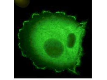
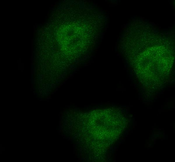
Immunofluorescence microscopy using Biorbyt's Monoclonal anti-HEF1 antibody (clone 2G9) shows detection of HEF1 localized at focal adhesion sites. The antibody was used at a 1:500 dilution with a 3-sec exposure time.
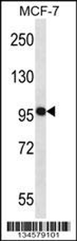
Western blot using Biorbyt's monoclonal anti-HEF1 antibody (clone 2G9) antibody shows detection of a ~92 kDa band corresponding to HEF1 in MCF7 lysate (p/n orb348664) [arrowhead]. Approximately 35 µg of lysate was loaded for SDS-PAGE followed by transfer onto nitrocellulose and reaction with a 1:1000 dilution of anti-HEF1 antibody. Detection occurred using a 1:5000 dilution of IRDye®800 conjugated Sh-a-Mouse IgG [H&L] for 45 min at room temperature (800 nm channel, green). Molecular weight estimation was made by comparison to prestained MW markers (indicated at left).
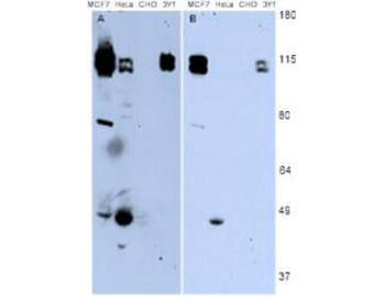
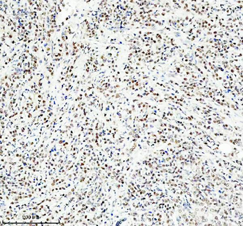
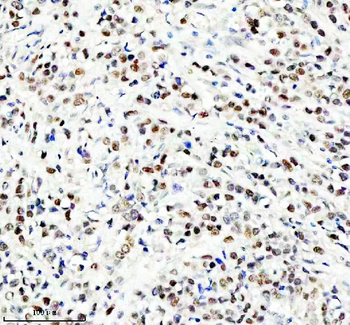
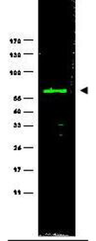
Western blotting using Biorbyt's monoclonal anti-HEF1 antibody (clone 2G9) shows detection of endogenous HEF1 present in various cell lines [MCF7, HeLa, CHO, 3Y1]. Panel A shows detection using a 15 min exposure. Panel B is the same blot exposed for 2 min. The doublet represents p105 and p115 staining. The lower MW band represents p55. 3Y1 cells are derived from rat fibroblast cells. Mouse 3T3 cells are also reactive (not shown). To date no staining has been noted on CHO cells.
Documents Download
Datasheet
Product Information
Request a Document
Protocol Information
WB
Western Blot (IB, immunoblot)
IF
Immunofluorescence
ELISA
Enzyme-linked Immunosorbent Assay (EIA)
IP
Immunoprecipitation
NEDD9 Antibody (orb344431)

- 0.0
Based on 0 reviews
Participating in our Biorbyt product reviews program enables you to support fellow scientists by sharing your firsthand experience with our products.
Login to Submit a ReviewAvailable Sizes
Select a size below
Choose Conjugation or Carrier Free Version
Free Secondary Antibody (20 ul)0/0
Please add an antibody product to your cart first.