You have no items in your shopping cart.
Description
Research Area
Epigenetics & Chromatin
Images & Validation
−Item 1 of 5
| Tested Applications | IF, IHC-P, IP, WB |
|---|---|
| Dilution Range | IF - 1:1,000, IP - 1:100, WB - 1:1000, IHC-P - 1:50-100 |
| Reactivity | Human |
| Predicted Reactivity | Mouse |
Key Properties
−| Host | Rabbit |
|---|---|
| Clonality | Polyclonal |
| Isotype | Rabbit IgG |
| Immunogen | This HDAC9 antibody is generated from rabbits immunized with a KLH conjugated synthetic peptide between 2-32 amino acids from the N-terminal region of human HDAC9. Antigen Region: 2-32 aa. |
| Target | HDAC9 |
| Molecular Weight | 111297 Da |
| Conjugation | Unconjugated |
Storage & Handling
−| Storage | Maintain refrigerated at 2-8°C for up to 2 weeks. For long term storage store at -20°C in small aliquots to prevent freeze-thaw cycles |
|---|---|
| Form/Appearance | Purified polyclonal antibody supplied in PBS with 0.09% (W/V) sodium azide. This antibody is prepared by Saturated Ammonium Sulfate (SAS) precipitation followed by dialysis against PBS. |
| Expiration Date | 12 months from date of receipt. |
| Disclaimer | For research use only |
Alternative Names
−Histone deacetylase 9, HD9, Histone deacetylase 7B, HD7, HD7b, Histone deacetylase-related protein, MEF2-interacting transcription repressor MITR, HDAC9, HDAC7, HDAC7B, HDRP, KIAA0744, MITR
Quality Guarantee
Explore bioreagents carefree to elevate your research. All our products are rigorously tested for performance. If a product does not perform as described on its datasheet, our scientific support team will provide expert troubleshooting, a prompt replacement, or a refund. For full details, please see our Terms & Conditions and Buying Guide. Contact us at [email protected].
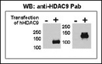
Both anti-HDAC9 N-term and C-term Pab were tested by WB and IP-WB using HeLa and HeLa-HDAC9 transfected cells. Top figure shows both Pab specifically detect HDAC9 in HeLa-HDAC9 transfected cell but not HeLa alone.
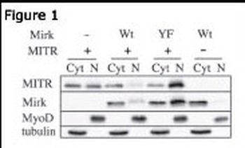
Immunoblots for MITR (HDAC9 N-term antibody), Mirk, MyoD and tubulin proteins are shown for cytoplasmic (Cyt) and nuclear (N) extracts from undifferentiated C2C12 myoblasts. Before cell collection for fractionation, the cells are transfected with plasmids coding for Mirk (Wt), kinase-inactive Mirk (YF) or MITR.
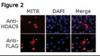
Figure 2: Immunofluorescence staining of MITR for a compartmentalization study in undifferentiated C2C12 myoblasts transfected with a MITR-expressing plasmid. MITR is detected by using the HDAC9 N-term antibody (top panel) or a FLAG antibody (bottom panel) detecting a FLAG epitope fused at the N-term end of the MITR construct.
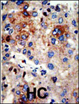
Formalin-fixed and paraffin-embedded human cancer tissue reacted with the primary antibody, which was peroxidase-conjugated to the secondary antibody, followed by AEC staining. This data demonstrates the use of this antibody for immunohistochemistry; clinical relevance has not been evaluated. BC = breast carcinoma; HC = hepatocarcinoma.

This figure shows that both Pab can immunoprecipitate (IP) HDAC9 from HeLa-HDAC9 tranfected cells.
Quick Database Links
Gene Symbol
HDAC9
UniProt
RefSeq (Protein):NP_848512.1, NP_848510.1, NP_001191075.1, NP_001191073.1, NP_055522.1, NP_001191076.1, NP_001191077.1, NP_001191074.1, NP_478056.1
UniProt Details
− No UniProt data available
NCBI Reference Sequences
−Associated Accession Numbers
Curated reference sequences for the gene transcript and protein productDocuments Download
Datasheet
Product Information
Request a Document
Protocol Information
WB
Western Blot (IB, immunoblot)
IHC-P
Immunohistochemistry Paraffin
IF
Immunofluorescence
IP
Immunoprecipitation
HDAC9 Antibody (N-term) (orb1938455)

- 0.0
Based on 0 reviews
Participating in our Biorbyt product reviews program enables you to support fellow scientists by sharing your firsthand experience with our products.
Login to Submit a ReviewAvailable Sizes
Select a size below
Choose Conjugation or Carrier Free Version
Free Secondary Antibody (20 ul)0/0
Please add an antibody product to your cart first.