You have no items in your shopping cart.
Description
Research Area
Signal Transduction
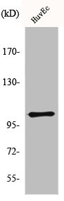
Images & Validation
−

Item 1 of 4
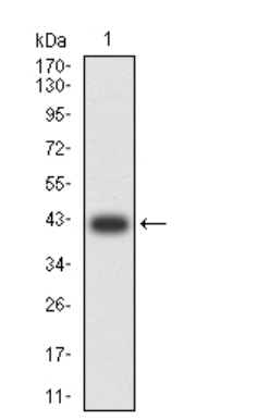
| Tested Applications | FC, ICC, WB |
|---|---|
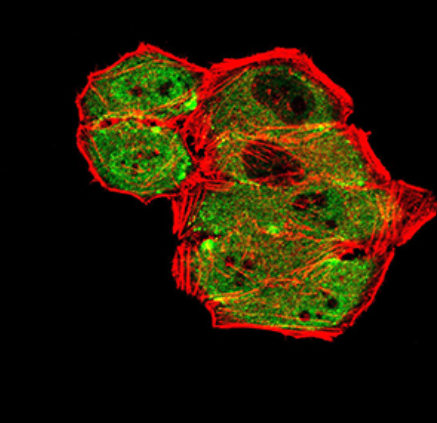
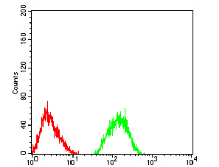
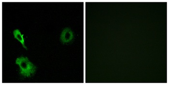
| Dilution Range | WB: 1:500-1:2,000, ICC: 1:50-1:200, FC: 1:50-1:100 |
| Reactivity | Human |
Key Properties
−| Antibody Type | Primary Antibody |
|---|---|
| Host | Mouse |
| Clonality | Polyclonal |
| Immunogen | Recombinant protein |
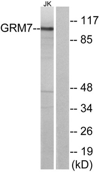
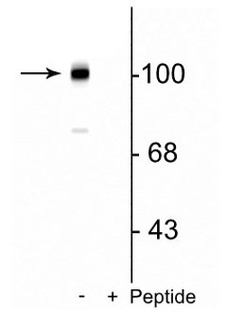
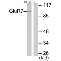
| Molecular Weight | 102 kDa |
| Purity | ProA affinity purified |
| Conjugation | Unconjugated |
Storage & Handling
−| Storage | Store at -20˚C |
|---|---|
| Form/Appearance | Liquid |
| Buffer/Preservatives | Formulation 1*TBS (pH7.4), 1%rAlbumin, 40%Glycerol. Preservative: 0.05% Sodium Azide |
| Expiration Date | 12 months from date of receipt. |
| Disclaimer | For research use only |
Similar Products
−- Item 1 of 4
mGluR7 rabbit pAb Antibody [orb768554]
ELISA, IHC, WB
Human, Mouse, Rat
Polyclonal
Unconjugated
50 μl, 100 μl - Item 1 of 1
- Item 1 of 1
Metabotropic Glutamate Receptor 7 (Ser862) Antibody [orb319578]
ELISA, WB
Bovine, Canine, Human, Primate, Zebrafish
Mouse, Rat
Rabbit
Polyclonal
Unconjugated
100 μl - Item 1 of 2
- Item 1 of 2
GRM7 Antibody [orb675707]
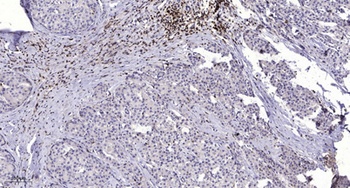
ELISA, IF, IHC, WB
Human, Mouse, Rat
Rabbit
Polyclonal
Unconjugated
100 μg, 50 μg


Quality Guarantee
Explore bioreagents carefree to elevate your research. All our products are rigorously tested for performance. If a product does not perform as described on its datasheet, our scientific support team will provide expert troubleshooting, a prompt replacement, or a refund. For full details, please see our Terms & Conditions and Buying Guide. Contact us at [email protected].
Quick Database Links
UniProt
UniProt Details
− No UniProt data available
Protocol Information
WB
Western Blot (IB, immunoblot)
FC
Flow Cytometry
ICC
Immunocytochemistry
Available Sizes
Select a size below
Choose Conjugation or Carrier Free Version
Free Secondary Antibody (20 ul)0/0
Please add an antibody product to your cart first.