You have no items in your shopping cart.
Description
Research Area
Stem Cell & Developmental Biology; Cell Biology
Images & Validation
−
Item 1 of 6
| Tested Applications | ELISA, FC, IF, IHC, WB |
|---|---|
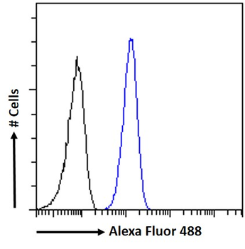
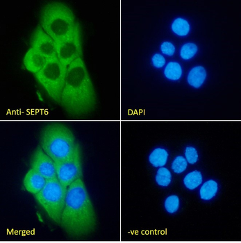
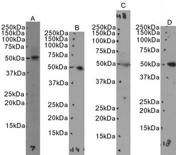
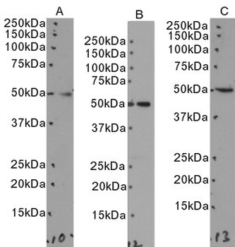
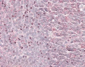
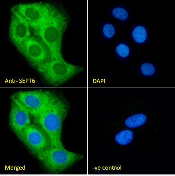
| Dilution Range | Peptide ELISA: antibody detection limit dilution 1:4000. Western blot: Approx 50kDa band observed in Human Testis, Duodenum, Kidney and Tonsil lysates, and in lysates of cell lines Daudi, Jurkat and MOLT-4 (calculated MW of 49.7kDa according to NP_055944.2). Recommended concentration: 0.1-1µg/ml. Primary incubation 1 hour at room temperature. IHC: In paraffin embedded Human Tonsil shows specific staining in dividing cells just outside a germinal centre. Recommended concentration: 3-5µg/ml. Immunofluorescence: Strong expression of the protein seen in A431 and U2OS cells. Recommended concentration: 10µg/ml. Flow Cytometry: Flow cytometric analysis of A431 cells. Recommended concentration: 10ug/ml. |
| Reactivity | Human |
| Predicted Reactivity | Bovine, Canine |
Key Properties
−| Clonality | Polyclonal |
|---|---|
| Immunogen | Peptide with sequence C-DEVNAFKQRKTA, from the internal region of the protein sequence according to NP_665798.1; NP_055944.2; NP_665801.1. |
| Target | SEPT6 |
| Protein Sequence | DEVNAFKQRKTA |
| Molecular Weight | 48.9 kDa; 49.7 kDa; 49.2 kDa |
| Purification | Purified from goat serum by ammonium sulphate precipitation followed by antigen affinity chromatography using the immunizing peptide. |
| Conjugation | Unconjugated |
Storage & Handling
−| Storage | Maintain refrigerated at 2-8°C for up to 2 weeks. For long term storage store at -20°C in small aliquots to prevent freeze-thaw cycles. |
|---|---|
| Buffer/Preservatives | Supplied at 0.5 mg/ml in Tris saline, 0.02% sodium azide, pH 7.3 with 0.5% bovine serum albumin. Aliquot and store at -20°C. Minimize freezing and thawing. |
| Expiration Date | 12 months from date of receipt. |
| Disclaimer | For research use only |
Alternative Names
−anti SEPT6 antibody, anti septin 6 antibody, anti KIAA0128 antibody, anti MGC16619 antibody, anti MGC20339 antibody, anti SEP2 antibody, anti SEPT2 antibody, anti septin 2 antibody
Similar Products
−- Item 1 of 1

Quality Guarantee
Explore bioreagents carefree to elevate your research. All our products are rigorously tested for performance. If a product does not perform as described on its datasheet, our scientific support team will provide expert troubleshooting, a prompt replacement, or a refund. For full details, please see our Terms & Conditions and Buying Guide. Contact us at [email protected].
Protocol Information
WB
Western Blot (IB, immunoblot)
IHC
Immunohistochemistry
FC
Flow Cytometry
IF
Immunofluorescence
ELISA
Enzyme-linked Immunosorbent Assay (EIA)
Available Sizes
Select a size below
Choose Conjugation or Carrier Free Version
Free Secondary Antibody (20 ul)0/0
Please add an antibody product to your cart first.