You have no items in your shopping cart.
Featured
Description
Research Area
Pharmacology & Drug Discovery
Images & Validation
−Item 1 of 3
| Tested Applications | ELISA, FC, IF, WB |
|---|---|
| Dilution Range | Peptide ELISA: antibody detection limit dilution 1:32000. Western blot: Approx 50kDa band observed in Human Tonsil lysates (calculated MW of 46.9kDa according to Human NP_001354640.1). Recommended concentration: 1-3µg/ml. Primary incubation 1 hour at room temperature. Immunofluorescence: Strong expression of the protein seen in the cytoplasm of HeLa cells. Recommended concentration: 10µg/ml. Flow Cytometry: Flow cytometric analysis of HeLa cells. Recommended concentration: 10ug/ml. |
| Reactivity | Human |
| Predicted Reactivity | Canine, Mouse, Rat |
Key Properties
−| Clonality | Polyclonal |
|---|---|
| Target | Histamine Receptor H2 |
| Protein Sequence | QEEKPLKLQVWSGTE |
| Molecular Weight | 44.5; 46.9 |
| Purification | Purified from goat serum by ammonium sulphate precipitation followed by antigen affinity chromatography using the immunizing peptide. |
| Conjugation | Unconjugated |
Storage & Handling
−| Storage | Maintain refrigerated at 2-8°C for up to 2 weeks. For long term storage store at -20°C in small aliquots to prevent freeze-thaw cycles. |
|---|---|
| Buffer/Preservatives | Supplied at 0.5 mg/ml in Tris saline, 0.02% sodium azide, pH 7.3 with 0.5% bovine serum albumin. Aliquot and store at -20°C. Minimize freezing and thawing. |
| Expiration Date | 12 months from date of receipt. |
| Disclaimer | For research use only |
Alternative Names
−anti HRH2 antibody, anti histamine receptor H2 antibody, anti HGNC:5183 antibody, anti H2R antibody, anti gastric receptor 1 antibody, anti OTTHUMP00000161242 antibody
Similar Products
−- Item 1 of 2
Histamine H2 Receptor rabbit pAb Antibody [orb768652]
ELISA, IF, WB
Human, Mouse, Rat
Polyclonal
Unconjugated
50 μl, 100 μl - Item 1 of 2
HRH2/Histamine H2 Receptor Antibody [orb1536930]
ICC, IF, IHC-P, WB
Human, Mouse, Rat
Rabbit
Polyclonal
Unconjugated
50 μl - Item 1 of 1
Goat anti-Histamine Receptor H2 (aa309-323) (mouse) Antibody [orb22498]
ELISA, WB
Mouse, Rat
Polyclonal
Unconjugated
100 μg - Item 1 of 2
HRH2 Rabbit Polyclonal Antibody [orb100935]
IF, IHC-Fr, IHC-P, WB
Canine, Rabbit, Rat
Human, Mouse
Rabbit
Polyclonal
Unconjugated
50 μl, 100 μl, 200 μl - Item 1 of 3


Quality Guarantee
Explore bioreagents carefree to elevate your research. All our products are rigorously tested for performance. If a product does not perform as described on its datasheet, our scientific support team will provide expert troubleshooting, a prompt replacement, or a refund. For full details, please see our Terms & Conditions and Buying Guide. Contact us at [email protected].
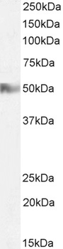
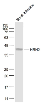
2 µg/ml staining of Human Tonsil lysate (35 µg protein in RIPA buffer). Detected by chemiluminescence.

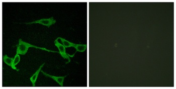
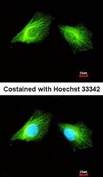
Immunofluorescence analysis of paraformaldehyde fixed HeLa cells, permeabilized with 0.15% Triton. Primary incubation 1hr (10 ug/ml) followed by Alexa Fluor 488 secondary antibody (2 ug/ml), showing cytoplasmic staining. The nuclear stain is DAPI (blue). Negative control: Unimmunized goat IgG (10 ug/ml) followed by Alexa Fluor 488 secondary antibody (2 ug/ml).
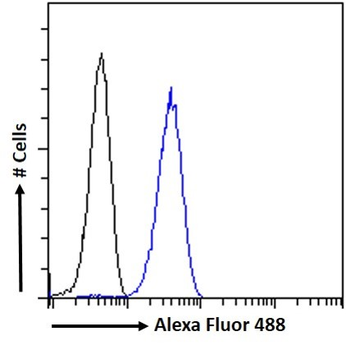
Flow cytometric analysis of paraformaldehyde fixed HeLa cells (blue line), permeabilized with 0.5% Triton. Primary incubation 1hr (10 ug/ml) followed by Alexa Fluor 488 secondary antibody (1 ug/ml). IgG control: Unimmunized goat IgG (black line) followed by Alexa Fluor 488 secondary antibody.
Documents Download
Datasheet
Product Information
Request a Document
Protocol Information
WB
Western Blot (IB, immunoblot)
FC
Flow Cytometry
IF
Immunofluorescence
ELISA
Enzyme-linked Immunosorbent Assay (EIA)
Filter by Species
Zhuang, Qian-Xing et al. Histamine Excites Striatal Dopamine D1 and D2 Receptor-Expressing Neurons via Postsynaptic H1 and H2 Receptors Mol Neurobiol, 55, 8059-8070 (2018)
Reactivity
Rat
Histamine Receptor H2 Antibody (orb19064)

- 0.0
Based on 0 reviews
Participating in our Biorbyt product reviews program enables you to support fellow scientists by sharing your firsthand experience with our products.
Login to Submit a ReviewAvailable Sizes
Select a size below
Choose Conjugation or Carrier Free Version
Free Secondary Antibody (20 ul)0/0
Please add an antibody product to your cart first.