You have no items in your shopping cart.
Description
Research Area
Immunology & Inflammation; Cell Biology
Images & Validation
−Item 1 of 3
| Tested Applications | ELISA, FC, IHC, WB |
|---|---|
| Dilution Range | Peptide ELISA: antibody detection limit dilution 1:128000. Western blot: This antibody was observed to recognize variations of bands at approx. 26kDa, 30kDa,35kDa and 70kDa in Human Duodenum and Lung lysates (calculated MW of 26.4kDa according to NP_004346.1 and 33.5kDa according to NP_001020330.1. All bands were blocked by incubation with the immunizing peptide and have been observed by other commercial sources. Recommended concentration: 0.5-1µg/ml. Primary incubation 1 hour at room temperature. IHC: Paraffin embedded Human Spleen. Recommended concentration: 5µg/ml Flow Cytometry: Flow cytometric analysis of Caco--2 cells. Recommended concentration: 10ug/ml. |
| Reactivity | Human |
Key Properties
−| Clonality | Polyclonal |
|---|---|
| Target | CD74 (aa159-171) |
| Protein Sequence | KGSFPENLRHLKN |
| Molecular Weight | 33.5; 26.4 |
| Purification | Purified from goat serum by ammonium sulphate precipitation followed by antigen affinity chromatography using the immunizing peptide. |
| Conjugation | Unconjugated |
Storage & Handling
−| Storage | Maintain refrigerated at 2-8°C for up to 2 weeks. For long term storage store at -20°C in small aliquots to prevent freeze-thaw cycles. |
|---|---|
| Buffer/Preservatives | Supplied at 0.5 mg/ml in Tris saline, 0.02% sodium azide, pH 7.3 with 0.5% bovine serum albumin. Aliquot and store at -20°C. Minimize freezing and thawing. |
| Expiration Date | 12 months from date of receipt. |
| Disclaimer | For research use only |
Alternative Names
−anti CD74 antibody, anti DHLAG antibody, anti HLADG antibody, anti Ia-GAMMA antibody, anti CD74 molecule, major histocompatibility complex, class II invariant chain antibody, anti CD74 antigen antibody, anti CD74 antigen (invariant polypeptide of major histocompatibility complex, class II antigen-associated) antibody, anti HLA-DR-gamma antibody, anti Ia-associated invariant chain antibody, anti MHC HLA-DR gamma chain antibody, anti gamma chain of class II antigens antibody
Quality Guarantee
Explore bioreagents carefree to elevate your research. All our products are rigorously tested for performance. If a product does not perform as described on its datasheet, our scientific support team will provide expert troubleshooting, a prompt replacement, or a refund. For full details, please see our Terms & Conditions and Buying Guide. Contact us at [email protected].
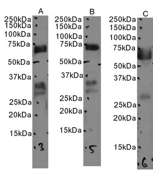
Primary incubation 1 hour at room temperature. Images A, B, C: Human Duodenum lysate, Human Lung lysate 1 and Human Lung lysate 2 at primary Ab concentration 1 µg/ml. (Loaded 35 µg protein in RIPA buffer, per lane). Detected by chemiluminescence.
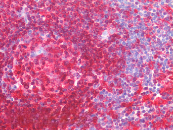
5 µg/ml staining of paraffin embedded Human Spleen. Steamed antigen retrieval with citrate buffer pH6, AP-staining.
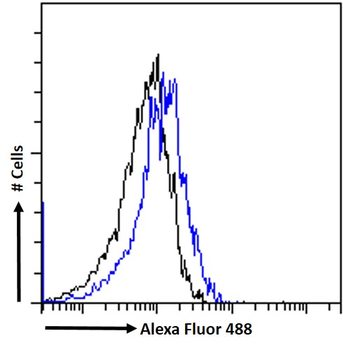
Flow cytometric analysis of paraformaldehyde fixed CaCo2 cells (blue line) permeabilized with 0.5% Triton. Primary incubation 1hr (10 ug/ml) followed by Alexa Fluor 488® conjugated secondary antibody (1 ug/ml). IgG control: Unimmunized goat IgG (black line) followed by Alexa Fluor 488 secondary antibody.
Documents Download
Datasheet
Product Information
Request a Document
Protocol Information
WB
Western Blot (IB, immunoblot)
IHC
Immunohistochemistry
FC
Flow Cytometry
ELISA
Enzyme-linked Immunosorbent Assay (EIA)
CD74 (aa159-171) Antibody (orb195359)

- 0.0
Based on 0 reviews
Participating in our Biorbyt product reviews program enables you to support fellow scientists by sharing your firsthand experience with our products.
Login to Submit a ReviewAvailable Sizes
Select a size below
Choose Conjugation or Carrier Free Version
Free Secondary Antibody (20 ul)0/0
Please add an antibody product to your cart first.