You have no items in your shopping cart.
Description
Images & Validation
−Item 1 of 7
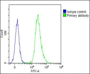
| Tested Applications | ELISA, IF, IHC, WB |
|---|---|
| Dilution Range | ELISA: 1:24,800-1:44,800, IHC: 1:100, IF: 5-15µg/mL |
| Reactivity | Human |
| Application Notes |
Key Properties
−| Antibody Type | Primary Antibody |
|---|---|
| Host | Rabbit |
| Clonality | Polyclonal |
| Immunogen | Glucagon antibody was prepared from whole rabbit serum produced by repeated immunizations with a synthetic peptide corresponding to an internal portion of human Glucagon. |
| Target | GCG |
| Purity | This affinity purified antibody is directed against human Glucagon. This product was affinity purified from monospecific antiserum by immunoaffinity purification. A BLAST analysis was used to suggest cross-reactivity with the antigen based on 100% homology with the immunizing sequence to human, chimpanzee, and bonobo. |
| Conjugation | Unconjugated |
Storage & Handling
−| Storage | Store vial at -20° C prior to opening. Aliquot contents and freeze at -20° C or below for extended storage. Avoid cycles of freezing and thawing. Centrifuge product if not completely clear after standing at room temperature. This product is stable for several weeks at 4° C as an undiluted liquid. Dilute only prior to immediate use. |
|---|---|
| Form/Appearance | Liquid (sterile filtered) |
| Buffer/Preservatives | Preservative: 0.01% (w/v) Sodium Azide. Stabilizer: None; Buffer: 0.02 M Potassium Phosphate, 0.15 M Sodium Chloride, pH 7.2 |
| Concentration | 1.18 mg/mL |
| Expiration Date | 12 months from date of receipt. |
| Dry Ice Shipping | Please note: This product requires shipment on dry ice. A dry ice surcharge will apply. |
| Disclaimer | For research use only |
Alternative Names
−Rabbit Anti-Glucagon, Pro-glucagon, Glicentin, Oxyntomodulin, OXM, OXY, Glucagon
Similar Products
−- Item 1 of 12
GLP-1 Mouse Monoclonal Antibody [orb10721]
IF, IHC-Fr, IHC-P
Mouse, Rat
Human, Mouse, Rat
Mouse
Monoclonal
Unconjugated
50 μl, 100 μl, 200 μl, 200 μg - Item 1 of 7
GLP2 Rabbit Polyclonal Antibody [orb10724]
ICC, IF, IHC-P, WB
Guinea pig, Human, Mouse, Porcine, Rat
Rabbit
Polyclonal
Unconjugated
100 μg - Item 1 of 6
Glucagon Antibody (C-term) [orb1929798]
FC, IF, IHC-P, WB
Human
Rabbit
Polyclonal
Unconjugated
50 μl, 100 μl - Item 1 of 3
GLP-1 (7-36) Rabbit Polyclonal Antibody [orb10719]
IF, IHC-Fr, IHC-P
Bovine, Porcine, Sheep
Human, Mouse, Rat
Rabbit
Polyclonal
Unconjugated
50 μl, 100 μl, 200 μl - Item 1 of 5
GLP1/GCG Rabbit Polyclonal Antibody [orb316568]
IF, IHC
Human, Mouse, Rat
Rabbit
Polyclonal
Unconjugated
100 μg


Quality Guarantee
Explore bioreagents carefree to elevate your research. All our products are rigorously tested for performance. If a product does not perform as described on its datasheet, our scientific support team will provide expert troubleshooting, a prompt replacement, or a refund. For full details, please see our Terms & Conditions and Buying Guide. Contact us at [email protected].
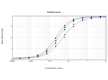
ELISA Results of Rabbit Anti-Glucagon Antibody. Each well was coated with 1 µg of conjugate. The starting concentration of antibody in the dilution series was 5 µg/ml. The titer is 1:34800 Glucagon - Free peptide [Green Line], 1:47200 Glucagon Standard - BSA conjugated [Blue Line], and 1:20500 Glucagon - BSA conjugated [Purple Line]. Each point on the Y-axis represents a 3-fold dilution. 3% Fish Gel (p/n orb348587), HRP conjugated Goat anti-Rabbit IgG (H&L) (p/n orb347673), and TMB substrate were used for detection.
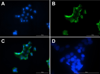
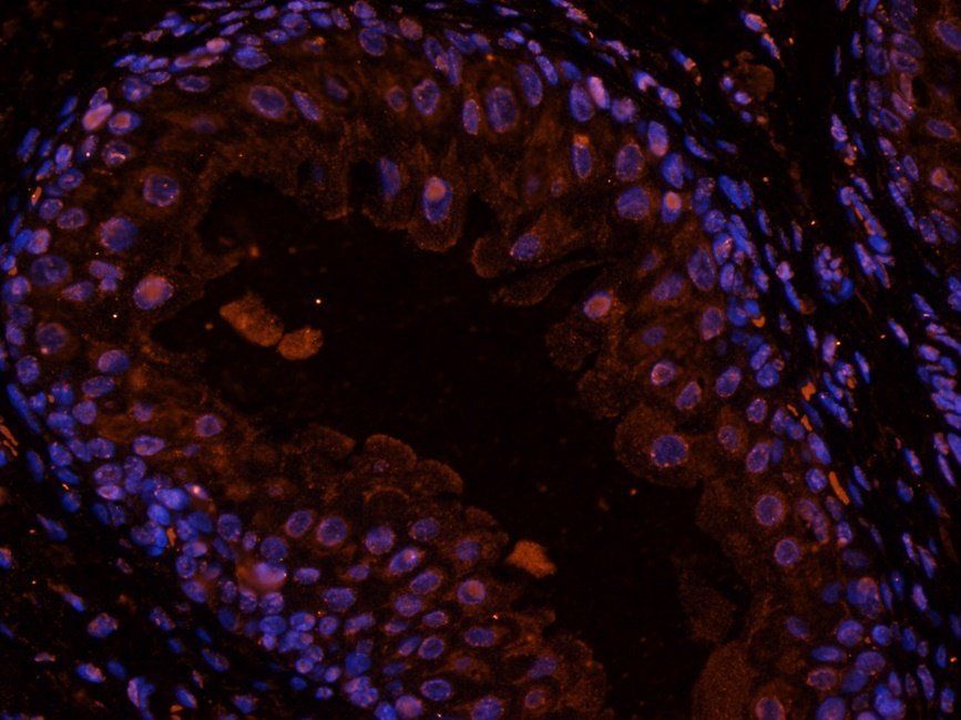
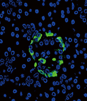
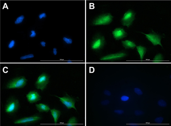
Immunofluorescence of Rabbit Anti-Glucagon Antibody. Cell Line: MCF7 cells. Fixative: 100% Methanol. Permeabilization: 0.3% Triton X-100. Primary Antibody: Anti-Glucagon at 15 µg/ml overnight at 2-8°C. Secondary Antibody: Goat Anti-Rabbit IgG DyLight™488 at 5 µl/mL for 1hr at RT. Nuclear Counterstain: DAPI. Staining: (A). DAPI. (B). Anti-Glucagon + DyLight™488 secondary. (C). Merge A + B. (D). secondary only. Localization expected: Cytoplasm.
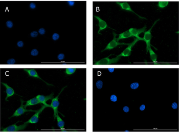
Immunofluorescence of Rabbit Anti-Glucagon Antibody. Cell Line: NIH/3T3 cells. Fixative: 100% Methanol. Permeabilization: Triton X-100. Primary Antibody: Anti-Glucagon at 15 µg/ml overnight at 2-8°C. Secondary Antibody: Goat Anti-Rabbit IgG DyLight™488 at 5 µl/mL for 1hr at RT. Nuclear Counterstain: DAPI. Staining: (A). DAPI. (B). Anti-Glucagon + DyLight™488 secondary. (C). Merge A + B. (D). secondary only. Localization expected: Cytoplasm.
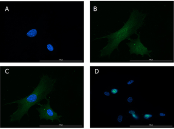
Immunofluorescence of Rabbit Anti-Glucagon Antibody. Cell Line: NIH/3T3 cells. Fixative: 4% PFA. Permeabilization: Triton X-100. Primary Antibody: Anti-Glucagon at 15 µg/ml overnight at 2-8°C. Secondary Antibody: Goat Anti-Rabbit IgG DyLight™488 at 5 µl/mL for 1hr at RT. Nuclear Counterstain: DAPI. Staining: (A). DAPI. (B). Anti-Glucagon + DyLight™488 secondary. (C). Merge A + B. (D). secondary only. Localization expected: Cytoplasm.
Immunofluorescence of Rabbit Anti-Glucagon Antibody. Cell Line: U20S cells. Fixative: 4% PFA. Permeabilization: 0.3% Triton X-100. Primary Antibody: Anti-Glucagon at 15 µg/ml overnight at 2-8°C. Secondary Antibody: Goat Anti-Rabbit IgG DyLight™488 at 5 µl/mL for 1hr at RT. Nuclear Counterstain: DAPI. Staining: (A). DAPI. (B). Anti-Glucagon + DyLight™488 secondary. (C). Merge A + B. (D). secondary only. Localization expected: Cytoplasm.
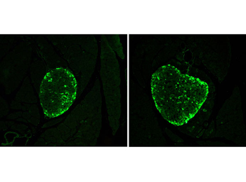
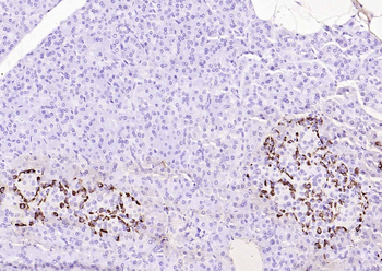
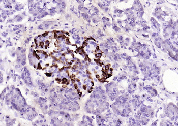
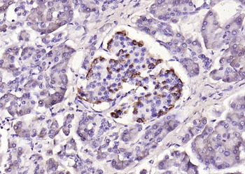
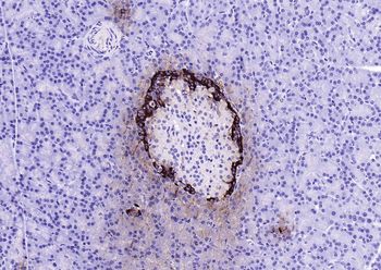
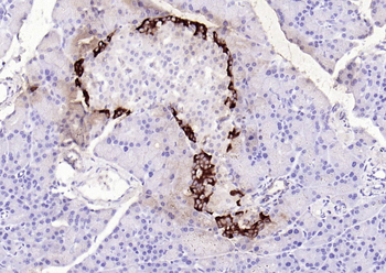
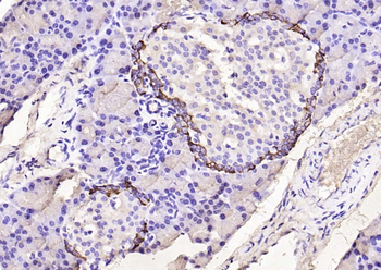
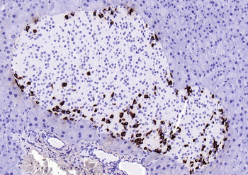
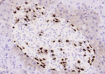
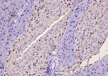
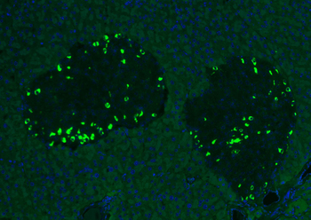
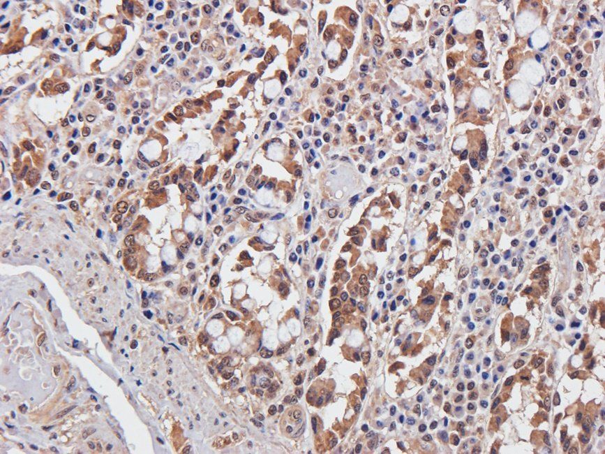
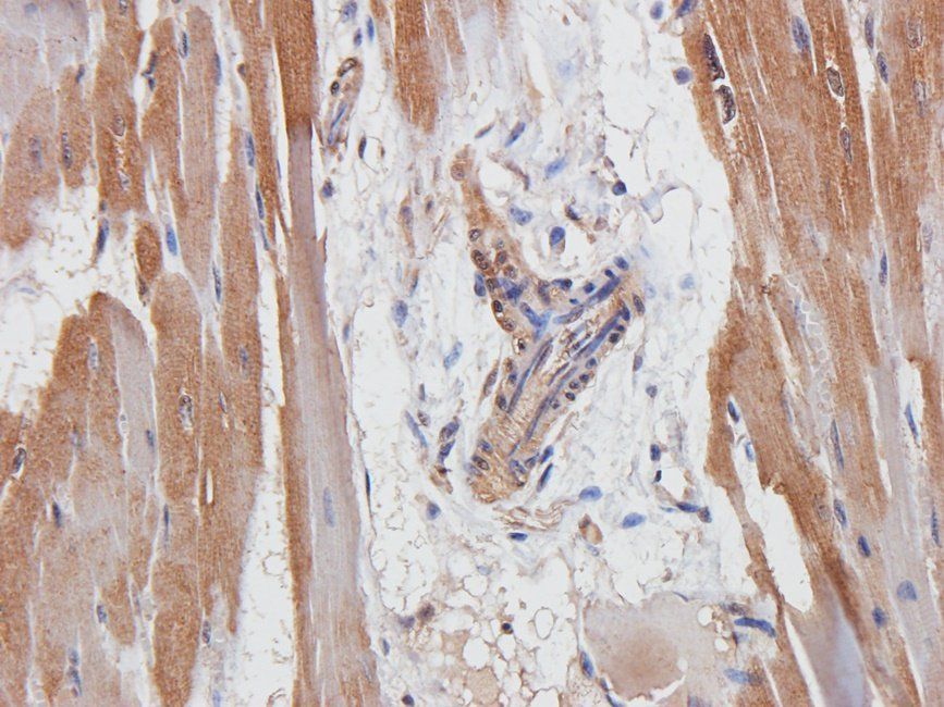
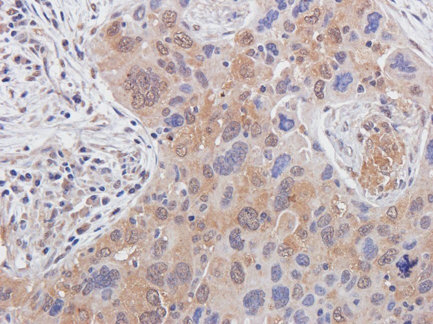
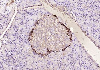
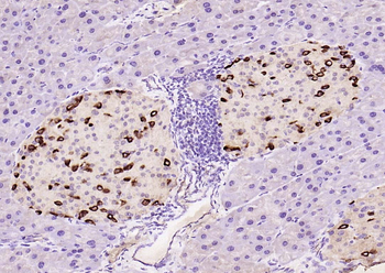
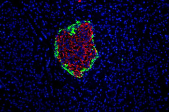
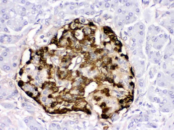
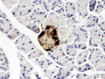
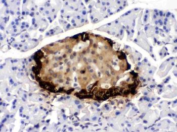
Immunohistochemistry results using Rabbit Anti-Glucagon Antibody. Tissue: alpha cells in CD1 mouse pancreatic islets. Fixation: 4% paraformaldehyde. Antigen Retrieval: 10 mM Sodium Citrate buffer for 10 mins at 95-100°C. Blocking: PBS, 1% ovalbumin, 0.3% Triton X-100. Primary Antibody: Anti-Glucagon at 1:100 overnight at RT. Secondary Antibody: Anti-Rabbit Alexa Fluor 488 at 1:500 for 1hr at RT. Original magnification 20x.
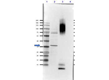
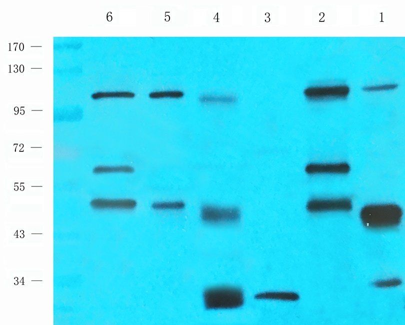
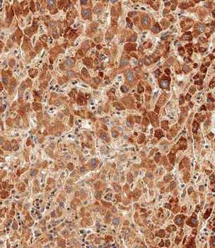
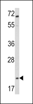
Western Blot of Rabbit Anti-Glucagon Antibody. Lane 1: Opal Prestained Molecular Weight. Lane 2: COS-7 Lysate - reduced (20 µg). Lane 3: BSA Conjugated Glucagon peptide - reduced (0.02 µg). Lane 4: Insulin - reduced (0.05 µg). Primary Antibody: Anti-Glucagon [Rabbit] Antibody at 1.0 µg/ml overnight at 2-8°C. Secondary Antibody: Goat Anti-Rabbit IgG (MX10) Peroxidase conjugated at 1:70000 for 30 mins at RT. Block: Blocking Buffer for Fluorescent Western Blotting (p/n orb348637) for 1hr at RT. Expected MW: ~21kDa. Observed MW: endogenous detection in COS-7 Lysate at ~35kDa. Glucagon peptide is detected at the MW of BSA. No cross-reactivity with insulin is observed. Exposure: 25 sec.
Quick Database Links
UniProt Details
− No UniProt data available
NCBI Reference Sequences
−Associated Accession Numbers
Curated reference sequences for the gene transcript and protein product| Protein | NP_002045.1 |
|---|
Documents Download
Datasheet
Product Information
Request a Document
Protocol Information
WB
Western Blot (IB, immunoblot)
IHC
Immunohistochemistry
IF
Immunofluorescence
ELISA
Enzyme-linked Immunosorbent Assay (EIA)
GCG Antibody (orb1784557)

- 0.0
Based on 0 reviews
Participating in our Biorbyt product reviews program enables you to support fellow scientists by sharing your firsthand experience with our products.
Login to Submit a ReviewAvailable Sizes
Select a size below
Choose Conjugation or Carrier Free Version
Free Secondary Antibody (20 ul)0/0
Please add an antibody product to your cart first.