You have no items in your shopping cart.
Featured
Description
Research Area
Neuroscience
Images & Validation
−Item 1 of 3
| Tested Applications | AM, ICC, IF, IHC, WB |
|---|---|
| Dilution Range | WB (1:1000), IHC-P (1:1000), ICC/IF (1:100) |
| Reactivity | Human, Mouse, Rat |
| Application Notes |
Key Properties
−| Host | Mouse |
|---|---|
| Clonality | Monoclonal |
| Isotype | IgG1 |
| Clone No. | N87/25 (Formerly sold as S87-25) |
| Immunogen | Fusion protein amino acids 370-433 of mouse GABA-A-R-Beta 3 |
| Target | GABA A Receptor |
| Molecular Weight | 55kDa |
| Purification | Protein G Purified |
| Conjugation | FITC |
Storage & Handling
−| Storage | Conjugated antibodies should be stored according to the product label |
|---|---|
| Buffer/Preservatives | 640.91mM DMSO, 136.36 mM Ethanolamine, 126.89 mM chlorides, 9.09mM phosphates, 9.09mM NaHCO3 |
| Concentration | 1 mg/ml |
| Expiration Date | 12 months from date of receipt. |
| Disclaimer | For research use only |
Alternative Names
−ECA5, GABA alpha receptor beta-2 subunit, GABA(A) receptor subunit beta-3, GABAA receptor beta 3 subunit, GABAA receptor subunit beta 3, GABR B3, GABRB3, Gamma aminobutyric acid (GABA) A receptor beta 3, Gamma aminobutyric acid receptor subunit beta 3, Gamma-aminobutyric acid receptor subunit beta-3, GBRB3_HUMAN, MGC9051
Similar Products
−- Item 1 of 3
GABA A Receptor Antibody (FITC) [orb182078]
AM, ICC, IF, IHC, WB
Human, Mouse, Rat
Mouse
Monoclonal
FITC
100 μg - Item 1 of 3
GABA A Receptor Antibody (FITC) [orb376793]
ICC, IF, IHC, WB
Human, Mouse, Rat
Mouse
Monoclonal
FITC
100 μg - Item 1 of 3
GABA A Receptor Antibody (FITC) [orb148893]
AM, ICC, IF, WB
Human, Mouse, Rat
Mouse
Monoclonal
FITC
100 μg - Item 1 of 2
GABA A Receptor Antibody (FITC) [orb376827]
ICC, IF, IHC, WB
Human, Mouse, Rat
Mouse
Monoclonal
FITC
100 μg - Item 1 of 2
GABA A Receptor Antibody (FITC) [orb376810]
ICC, IF, IHC, WB
Human, Mouse, Rat
Mouse
Monoclonal
FITC
100 μg


Quality Guarantee
Explore bioreagents carefree to elevate your research. All our products are rigorously tested for performance. If a product does not perform as described on its datasheet, our scientific support team will provide expert troubleshooting, a prompt replacement, or a refund. For full details, please see our Terms & Conditions and Buying Guide. Contact us at [email protected].
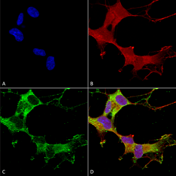
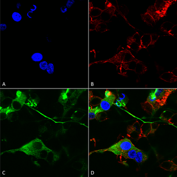
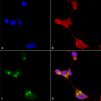
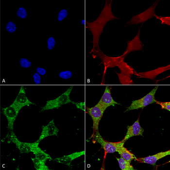
Immunocytochemistry/Immunofluorescence analysis using Mouse Anti-GABA-A Receptor Beta 3 Monoclonal Antibody, Clone N87/25. Tissue: Neuroblastoma cells (SH-SY5Y). Species: Human. Fixation: 4% PFA for 15 min. Primary Antibody: Mouse Anti-GABA-A Receptor Beta 3 Monoclonal Antibody at 1:100 for overnight at 4°C with slow rocking. Secondary Antibody: AlexaFluor 488 at 1:1000 for 1 hour at RT. Counterstain: Phalloidin-iFluor 647 (red) F-Actin stain; Hoechst (blue) nuclear stain at 1:800, 1.6mM for 20 min at RT. (A) Hoechst (blue) nuclear stain. (B) Phalloidin-iFluor 647 (red) F-Actin stain. (C) GABA-A Receptor Beta 3 Antibody (D) Composite.
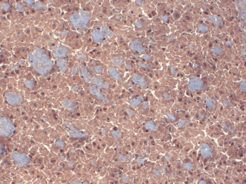
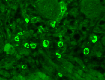
Immunohistochemistry analysis using Mouse Anti-GABA A Receptor Monoclonal Antibody, Clone N87/25. Tissue: Brain. Species: mouse. Fixation: 10% Formalin Solution for 12-24 hours at RT. Primary Antibody: Mouse Anti-GABA A Receptor Monoclonal Antibody at 1:1000 for 1 hour at RT. Secondary Antibody: HRP/DAB Detection System: Biotinylated Goat Anti-Mouse, Streptavidin Peroxidase, DAB Chromogen (brown) for 30 minutes at RT. Counterstain: Mayer Hematoxylin (purple/blue) nuclear stain at 250-500 μl for 5 minutes at RT.
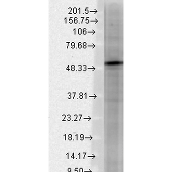
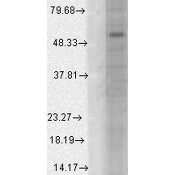
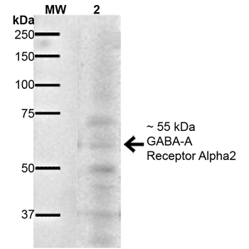
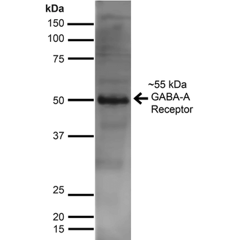
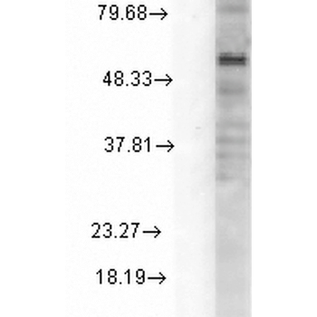
Western Blot analysis of Rat brain membrane lysate showing detection of GABA A Receptor protein using Mouse Anti-GABA A Receptor Monoclonal Antibody, Clone N87/25. Load: 15 μg. Block: 1.5% BSA for 30 minutes at RT. Primary Antibody: Mouse Anti-GABA A Receptor Monoclonal Antibody at 1:1000 for 2 hours at RT. Secondary Antibody: Sheep Anti-Mouse IgG: HRP for 1 hour at RT.
Quick Database Links
UniProt Details
− No UniProt data available
NCBI Gene Details
− No NCBI Gene data available
NCBI Reference Sequences
−Associated Accession Numbers
Curated reference sequences for the gene transcript and protein product| Protein | NP_032097.1 |
|---|
Documents Download
Datasheet
Product Information
Request a Document
Protocol Information
WB
Western Blot (IB, immunoblot)
IHC
Immunohistochemistry
IF
Immunofluorescence
ICC
Immunocytochemistry
GABA A Receptor Antibody (FITC) (orb181747)

- 0.0
Based on 0 reviews
Participating in our Biorbyt product reviews program enables you to support fellow scientists by sharing your firsthand experience with our products.
Login to Submit a ReviewAvailable Sizes
Select a size below
Choose Conjugation or Carrier Free Version
Free Secondary Antibody (20 ul)0/0
Please add an antibody product to your cart first.